32 Disorders of the Eye
General pediatricians must be able to identify common ocular problems in infants and children. Many of these conditions can be managed by the pediatrician, but others require referral to an ophthalmologist. Evaluation of the eye and adnexa involves inspection of the eyelids and preauricular lymph nodes; the extraocular movements, confrontational fields, and pupils; red light reflex in young children and fundus examination if indicated; and the globe itself, noting the conjunctiva, cornea, and sclera. Visual acuity can be assessed using several instruments, most commonly the Snellen eye chart. Corneal epithelial defects can be detected by applying fluorescein dye and then illuminating the cornea with a blue-filtered light or Wood’s lamp. Further evaluation by slit lamp and tonometry can be performed by an ophthalmologist if required.
Abnormal Red Light Reflex
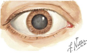
All children should have an examination of the red reflex within the first 2 months of life. Children with dark spots in the red reflex, a blunted or absent red reflex, or a white reflex (leukocoria) should be referred to an ophthalmologist. An abnormal red reflex can result from corneal opacities, aqueous opacities, vitreous opacities, and retinal lesions. Leukocoria may indicate pathology, including metabolic, inflammatory, infectious, toxic, oncologic, and traumatic causes; the most common are congenital cataracts and retinoblastoma.
Cataracts
Congenital cataracts occur in two in 10,000 births (Figure 32-1). Of these, 20% to 25% of cases occur secondary to a congenital infection (rubella, cytomegalovirus, or toxoplasmosis) or as a component of a genetic or metabolic condition, such as Turner syndrome, Down syndrome, trisomy 13 and 18, galactosemia, and peroxisomal disorders. Children exposed to high-dose long-term corticosteroid therapy are also at risk, as are children with uveitis or who sustain ocular trauma.
Retinoblastoma
Retinoblastoma occurs in one in 15,000 live births, and 250 to 300 new cases are diagnosed annually in the United States. The hereditary form is caused by a mutation in the retinoblastoma gene, a tumor suppressor gene. A second mutation is then necessary for tumor growth. In the hereditary form, approximately 60% of cases are bilateral and are associated with other cancers, notably osteosarcoma. Retinoblastoma can present with leukocoria and strabismus, and in more advanced cases, proptosis, eye pain, or hyphema. The extent of the disease should be evaluated by computed tomography (CT) or magnetic resonance imaging and orbital ultrasonography. The average age at diagnosis is 1 year for bilateral cases and 2 years for unilateral cases. Urgent referral to an ophthalmologist for potentially vision-sparing and lifesaving treatment is imperative.
Disorders of Eye Movement
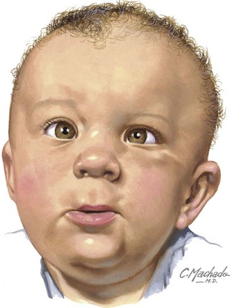
Strabismus
Misalignment of the eyes affects approximately 4% of children younger than 6 years of age (Figure 32-2). Heterophoria is the intermittent tendency for eyes to deviate, and heterotropia is a constant misalignment. The prefixes eso- (inward), exo- (outward), hyper- (upward), and hypo- (downward) indicate the direction of the misaligned eye. Other causes of eye deviations are cranial nerve palsies, intracranial or intraorbital mass, increased intracranial pressure (ICP), and myasthenia gravis.
Heterophorias are usually not apparent; however, under certain conditions such as stress, fatigue, or illness, this latent deviation can be detected. If the deviation is large, patients may experience double vision (diplopia), headache, or eye strain. Heterotropias are present at all times.
Tropias can be tested using the corneal light reflex. The examiner shines a light onto both cornea and notes the placement of the light reflex. If strabismus is present, the reflected light is asymmetric on the cornea. To further test for strabismus, the examiner can perform a cover test. The child should look at an object in the distance. The examiner covers one eye and watches for movement in the uncovered eye. If movement occurs in the uncovered eye, then a misalignment exists in that eye. Phorias can be detected by covering the affected eye; when the eye is uncovered, the practitioner will note the eye moving back into alignment.
Early detection of strabismus is essential because amblyopia can develop if misalignment persists, resulting in permanent visual impairment. Strabismus that is constant or intermittent strabismus that does not correct by age 3 months should prompt ophthalmology referral to begin treatment. The unaffected eye is patched or blurred (with glasses or drops), thereby forcing the strabismic eye to provide a retinal image to the brain and stimulate the proper visual development. In some cases, surgery on the extraocular muscles is necessary to achieve proper alignment.
Amblyopia
Amblyopia is vision impairment caused by an interference with a clear retinal image in one or both eyes during the development of visual acuity in infancy and early childhood. Amblyopia occurs during the critical period of development before the cortex has become visually mature, mainly within the first decade of life. It can be caused by a deviated eye (strabismus), refractive error, or opacity within the visual axis. Treatment is specific to the cause such as strabismus correction, corrective glasses, or removal of the opacity.
Red Eye
Red eye is a common pediatric complaint. The differential diagnosis is broad, including infectious, allergic, and inflammatory causes, as well as trauma, glaucoma, and Kawasaki disease. The most common cause is conjunctivitis, inflammation of the conjunctivae, the mucous membrane that covers the surface of the eye up to the limbus (the junction of the sclera and the cornea) and continues onto the inside surface of the eyelids.
Viral Conjunctivitis
Viral conjunctivitis presents with watery or mucopurulent discharge, eye irritation, and scleral injection (Figure 32-3). Both eyes are usually affected simultaneously or in sequence. More serious infection causes pseudomembranes (inflammatory debris and fibrin) or true conjunctival membranes. Punctate keratitis and subepithelial opacities may also occur, causing decreased vision, photosensitivity, or glare and haloes around bright lights.
Adenovirus is the most common pathogen causing viral conjunctivitis. Other symptoms include fever, pharyngitis, rhinitis, cough, and preauricular lymphadenopathy. This infection is highly contagious, and those who are infected should avoid sharing towels and touching their eyes and should wash their hands frequently. Treatment is symptomatic, including cool compresses or antiallergy drops for itchiness. Topical antibiotics should be used if large epithelial defects are present.
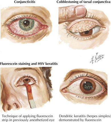
Other agents such as measles (rare because of widespread immunization), influenza, enterovirus, and herpes simplex virus (HSV) can cause conjunctivitis. Primary or recurrent HSV can also cause keratitis (corneal inflammation) with a dendrite pattern seen on fluorescein staining (see Figure 32-3). Treatment of ocular HSV includes topical antivirals (trifluridine, vidarabine, or iododeoxyuridine) and, depending on the extent of infection, oral or intravenous acyclovir. Consultation with an ophthalmologist is recommended.

Stay updated, free articles. Join our Telegram channel

Full access? Get Clinical Tree

