50 Disorders of Red Blood Cells
Red blood cells (RBC) are non-nucleated cells composed of a cell membrane, complex surface glycoproteins, and hemoglobin (Hb). Hb, the major component of RBCs, facilitates oxygen transport from the lungs to tissue capillaries by reversible binding and releasing oxygen, according to the characteristics of the oxyhemoglobin dissociation curve. As a result, RBC homeostasis is essential to prevent tissue hypoxia and maintain critical organ function.
RBC disorders can be divided into two categories, congenital and acquired disorders. Congenital disorders include membrane defects, thalassemia, hemoglobinopathies, and enzyme defects and aplasia. Acquired disorders include immune destruction, mechanical destruction, anemia of chronic disease, nutritional deficiencies (i.e., deficiency of B12, folate, iron), and aplasia. These disorders have different clinical features and mechanisms of disease, but all result in anemia.
Etiology and Pathogenesis
Congenital Red Blood Cell Disorders
Membrane Defects
Hereditary spherocytosis (HS) is the most common RBC membrane defect, affecting one in 5000 people of Northern European desent. Two-thirds of cases are inherited by autosomal dominant transmission, but de novo mutations can occur. The disorder arises from abnormalities of RBC membrane proteins. Commonly affected membrane proteins include ankyrin, band 3, and spectrin. These abnormalities lead to an unstable RBC membrane that assumes a spherical shape rather than the biconcave disc shape found in normal RBCs. The spherical RBCs have less deformability and cannot circulate freely through narrow capillaries. As a result, they become trapped in the spleen and are engulfed by macrophages, leading to a shortened RBC lifespan.
Hereditary elliptocytosis (HE) is another RBC membrane defect characterized by elliptical or oval-shaped RBCs on the peripheral blood smear. Unlike HS, HE is more common in people of African and Mediterranean ancestry. It is inherited mostly in an autosomal dominant fashion, although there can be spontaneous mutations. Clinical manifestations are usually similar to HS, but different phenotypic variations exist.
Thalassemias
The predominant adult hemoglobin A molecule is a tetramer formed by two α-globin and two β-globin chains. The thalassemias are a heterogeneous group of inherited disorders in which the production of normal Hb is partly or completely suppressed from the defective synthesis of one of the two globin chains (α or β). The type of thalassemia refers to the specific globin chain that is underproduced and is identified as either α- or β-thalassemia. A decrease in the production of either an α- or β-globin chain results in an excess of free globin chains that precipitate in the RBC and cause RBC membrane damage. The end result is anemia from RBC hemolysis and ineffective erythropoiesis in the bone marrow.
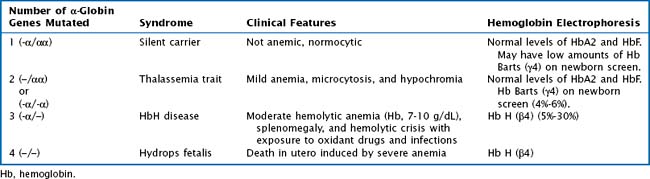
Whereas α -thalassemia is more commonly found in Southeast Asia, β-thalassemia is more common in Mediterranean countries. There are two α-globin genes located on chromosome 16. α-Thalassemias are usually the result of large gene deletions, causing a reduction in α-globin production. The severity of disease is directly related to the number of genes involved (Table 50-1).
There is one β-globin gene located on chromosome 11. Point mutations are the most common type of genetic mutation in β-thalassemia. The β–thalassemia trait occurs when only one gene is affected, resulting in a mild microcytic anemia. The Hb electrophoresis reveals an increased Hb A2 or Hb F level. In contrast the inheritance of two affected β-globin genes results in a broad spectrum of clinical disease. The severity is determined by the residual amount of β-globin synthesis. The clinical phenotype ranges from transfusion dependence (thalassemia major) to a moderate anemia that does not necessitate chronic transfusions (thalassemia intermedia). Severe β-thalassemia is diagnosed between 6 months and 2 years of age. Laboratory analysis reveals a moderate to severe microcytic anemia and 20% to 100% HbF, 2% to 7% HbA2, and 0% to 80% HbA. Clinically, patients present with pallor, failure to thrive, hepatosplenomegaly, and bone deformities from marrow expansion.
Hemoglobinopathies
The hemoglobinopathies are a group of autosomal recessive inherited disorders characterized by synthesis of abnormal Hb molecules (i.e., S, and C). The most common and severe hemoglobinopathy is sickle cell disease, in which only HbS is produced. Chapter 53 of this book is dedicated to the in-depth discussion of sickle cell disease.
Enzyme Deficiencies
Glucose-6-phosphate dehydrogenase (G6PD) deficiency is an x-linked recessive disorder characterized by abnormally low levels of the enzyme G6PD. Worldwide it is the most common enzyme deficiency. G6PD is the rate-limiting enzyme of the pentose phosphate pathway, which is crucial for protecting RBCs from oxidative stress. In G6PD deficiency, damage by oxidant free radicals causes RBCs to hemolyze.
The severity of disease is based on the baseline G6PD level. The majority of individuals with G6PD have a moderate deficiency (10% normal activity), and at a steady state, they are hematologically normal. But with exposure to an oxidative stressor, they can develop acute hemolysis with resultant anemia, reticulocytosis, and hyperbilirubinemia or jaundice. Patients with a severe G6PD deficiency (i.e., Mediterranean variant) can have baseline mild anemia and reticulocytosis. Drugs that are oxidative stressors and should be avoided in patients with G6PD include antimalarials, sulfonamides and sulfones, quinolones, aspirin, methylene blue, and rasburicase. Other categories of oxidative stressors to avoid include fava beans and naphthalene mothballs. The degree of hemolysis varies with the drug’s antioxidant effect, the amount ingested, and the severity of the enzyme deficiency in the patient. The highest prevalence of disease is among persons of African, Asian, and Mediterranean descent.
Pyruvate kinase (PK) deficiency is an inherited metabolic disorder of the enzyme PK, which catalyzes the rate-limiting step in the glycolysis pathway. A deficiency of the enzyme PK compromises RBC adenosine triphosphate production and metabolic energy demand, leading to hemolysis. The inheritance pattern is usually autosomal recessive. Clinically, patients have a moderate to severe hemolytic anemia, reticulocytosis (may be 40%-70%), jaundice, and splenomegaly. Symptoms caused by hemolysis range from mild to severe.
Diamond-Blackfan Anemia
Diamond Blackfan anemia (DBA) is a congenital, lifelong pure RBC aplasia characterized by anemia, reticulocytopenia, and normocellular bone marrow with a paucity of erythroid precursors. The white blood cell count and platelets are usually normal. The anemia can be mildly macrocytic or normocytic and usually manifests after birth or in the first few months of life. DBA is associated with various congenital abnormalities, most commonly short stature, craniofacial abnormalities, and thumb abnormalities (classically triphalangeal thumbs). It is also associated with an increased predisposition to cancer. Inheritance patterns vary, since dominant, recessive, and sporadic mutations can all cause DBA. Approximately 50% of patients with DBA have a single mutation in a gene encoding for a ribosomal protein.
Congenital Bone Marrow Failure Syndromes
Inherited bone marrow failure syndromes include Fanconi anemia, dyskeratosis congenita, Schwachman-Diamond syndrome, Pearson syndrome, and amegakaryocytic thrombocytopenia. In contrast to DBA, these disorders affect multiple cell lines. These disorders are rare. Presentation can occur at a young age with symptoms of pancytopenia and congenital malformations, which differ by syndrome.
Acquired Red Blood Cell Disorders
Mechanical Red Blood Cell Destruction
The previous congenital RBC disorders are caused by abnormalities inherent to RBCs. In contrast, mechanical destruction involves hemolysis caused by extrinsic factors unrelated to the RBCs. Examples of mechanical RBC destruction include vascular lesions (i.e., AVMs), cardiac valvular defects, and microangiopathic damage (i.e., hemolytic uremic syndrome or thrombotic thrombocytopenic purpura). RBCs can also be destroyed by infections, drugs, toxins, and heat (with severe burns).
Autoimmune Hemolytic Anemia
The most common immune-mediated extrinsic anemia is autoimmune hemolytic anemia (AIHA), in which circulating antibodies are directed against the patient’s RBCs. AIHA can be primary or secondary to infection, drugs, or an underlying disease process such as lymphoma, systemic lupus erythematosus, or immunodeficiency. Primary AIHA occurs in the majority of children and often occurs after a viral illness. The specific autoantibodies can be either IgG in warm-reactive antibodies or IgM in cold-agglutinin disease. Patients with AIHA require close monitoring because brisk hemolysis can result in a sudden decrease in Hb that can be life threatening.

Stay updated, free articles. Join our Telegram channel

Full access? Get Clinical Tree

