Disorders of Phagocyte Function

Chapter 124 Disorders of Phagocyte Function


Neutrophils are particularly important in protecting the skin, the lining of the respiratory and gastrointestinal tracts, and other mucous membranes as part of the 1st line of defense against microbial invasion. During the critical 2-4 hr after microbial invasion, these phagocytes arrive at the site of inflammation to contain the infection and prevent hematogenous dissemination.


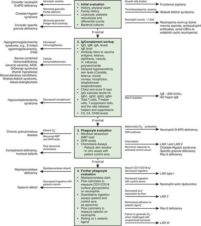
Immunologic evaluation of patients with suspected immunodeficiency (Chapter 116) should focus on disorders of phagocyte function (Table 124-1) in patients with recurrent or unusual bacterial infections (Fig. 124-1).


Chemotaxis, the direct migration of cells into sites of infection, involves a complex series of events (Chapter 121). Studies of defective in vitro chemotaxis of neutrophils obtained from children having various clinical conditions have not established whether frequent infections arise from a primary chemotactic abnormality or occur as secondary medical complications of the underlying disorder. For example, variable but at times severe abnormalities in neutrophil motility accompany the hyper-IgE syndrome, which is characterized by markedly elevated levels of IgE, chronic dermatitis, and recurrent sinopulmonary infections, as well as coarse facial features, retention of primary teeth, and a propensity for recurrent bone fractures (Chapter 123).


Leukocyte Adhesion Deficiency


Leukocyte adhesion deficiency 1 (LAD-1), 2 (LAD-2), and 3 (LAD-3) are rare autosomal recessive disorders of leukocyte function. LAD-1 affects about 1 per 10 million individuals and is characterized by recurrent bacterial and fungal infections and depressed inflammatory responses despite striking blood neutrophilia.


Genetics and Pathogenesis


LAD-1 results from mutations of the gene on chromosome 21q22.3 encoding CD18, the 95-kD β2 leukocyte integrin subunit. Normal neutrophils express 4 heterodimeric adhesion molecules: LFA-1 (CD11a/CD18), Mac-1 (CD11b/CD18, also known as CR3 or iC3b receptor), p150,95 (CD11c/CD18), and αd β2 (CD11d/CD18). These 4 transmembrane adhesion molecules are composed of unique α1 subunits of 185 kD, 190 kD, 150 kD, and 160 kD, respectively, encoded on chromosome 16, and share a common β2 subunit. This group of leukocyte integrins is responsible for the tight adhesion of neutrophils to the endothelial cell surface, egress from the circulation, and adhesion to iC3b-coated microorganisms, which promotes phagocytosis and particulate activation of the phagocyte NADPH oxidase.


Mutations in the CD18 gene either impair gene expression or affect the structure of the synthesized CD18 peptide, leading to functionally abnormal CD11/CD18. Some mutations of CD11/CD18 allow a low level of assembly and activity of integrin molecules. These children retain some neutrophil integrin adhesion function and have a moderate phenotype. Failure of neutrophils to bear the β2-integrins leads to inability to migrate to sites of inflammation outside the blood vessel lumen because of their inability to adhere firmly to surfaces and undergo transendothelial migration. Failure of the CD11/CD18–deficient neutrophils to undergo transendothelial migration occurs because the β2-integrins bind to intercellular adhesion molecules 1 (ICAM-1) and 2 (ICAM-2) expressed on inflamed endothelial cells (Chapter 121). Neutrophils that do arrive at inflammatory sites by CD11/CD18–independent processes fail to recognize microorganisms opsonized with complement fragment iC3b, an important stable opsonin formed by the cleavage of C3b. Hence, other neutrophil functions such as degranulation and oxidative metabolism normally triggered by iC3b binding are also markedly compromised in LAD-1 neutrophils, resulting in impaired phagocytic function and high risk for serious and recurrent bacterial infections.


Monocyte function is also impaired, with poor fibrinogen-binding function, an activity that is promoted by the CD11/CD18 complex. Consequently, such cells are unable to participate effectively in wound healing.


Children with LAD-2 share the clinical features of LAD-1 but have normal CD11/CD18 integrins. Features unique to LAD-2 include neurologic defects, cranial facial dysmorphism, and absence of the erythrocyte ABO blood group antigen (Bombay phenotype). LAD-2 (also known as congenital disorder of glycosylation IIc) derives from mutations in the gene encoding a specific GDP-L-fucose transporter of the Golgi apparatus. This abnormality prevents the incorporation of fucose into various cell surface glycoproteins, which are expressed on cell surface membranes. These include the erythrocyte carbohydrate blood group markers and neutrophil the carbohydrate structure sialyl Lewis X. Absence of this selectin ligand renders the cells incapable of rolling adhesion to activated endothelial cells, an initial step necessary for subsequent integrin-mediated activation, spreading, and transendothelial migration. Infections in LAD-2 are milder than that in LAD-1.


LAD-3 is characterized by a Glanzmann thrombasthenia-like bleeding disorder, delayed separation of the umbilical cord as well as serious skin and soft tissue infections similar to that seen in LAD-1, and failure of leukocytes to undergo β2 and β1 integrin mediated adhesion and migration. Mutations in KINDLIN3 affect integrin activation.


Clinical Manifestations


Patients with the severe clinical form of LAD-1 express <0.3% of the normal amount of the β2-integrin molecules, whereas patients with the moderate phenotype may express 2-7% of the normal amount. Children with severe disease present in infancy with recurrent, indolent bacterial infections of the skin, mouth, respiratory tract, lower intestinal tract, and genital mucosa. They may have a history of delayed separation of the umbilical cord, usually with associated infection (omphalitis) of the cord stump. However, 10% of healthy infants can have cord separation at age 3 wk or later, so this sign alone should not be sufficient to raise suspicion of LAD-1. Skin infection may progress to large chronic ulcers with polymicrobial infection, including anaerobic organisms. The ulcers heal slowly, need months of antibiotic treatment, and often require plastic surgical grafting. Severe gingivitis can lead to early loss of primary and secondary teeth.


The pathogens infecting patients with LAD-1 are similar to those affecting patients with severe neutropenia (Chapter 125) and include Staphylococcus aureus and enteric gram-negative organisms such as Escherichia coli. These patients are also susceptible to opportunistic infection by fungi such as Candida and Aspergillus. Typical signs of inflammation such as swelling, erythema, and warmth may be absent. Pus does not form, and few neutrophils are identified microscopically in biopsy specimens of infected tissues. Despite the paucity of neutrophils within the affected tissue, the circulating neutrophil count during infection typically exceeds 30,000/µL and can surpass 100,000/µL. During intervals between infections, the peripheral blood neutrophil count may chronically exceed 12,000/µL. LAD-1 genotypes producing moderate amounts of functional integrins at the surface of the neutrophil significantly reduce the severity and frequency of infections compared to children with the severe form.
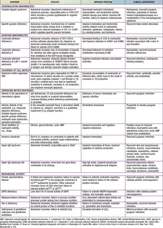
Similar clinical syndromes have been reported in patients with endothelial cell E-selectin deficiency, which manifests with delayed separation of the umbilical cord and omphalitis, and in a child with an autosomal dominant mutation of Rac2 (a Rho GTPase needed to regulate actin organization and superoxide production). Rac2 deficiency is characterized by delayed separation of the umbilical cord, leukocytosis, and absence of pus at sites of infection.


Jun 18, 2016 | Posted by in PEDIATRICS | Comments Off on Disorders of Phagocyte Function

Full access? Get Clinical Tree

Get Clinical Tree app for offline access