Disorders of Calcium and Bone Metabolism

69 Disorders of Calcium and Bone Metabolism


Physiological concentrations of plasma calcium and phosphorus are necessary to ensure skeletal integrity and to maintain vital physiological processes, including muscle contraction, coagulation, energy metabolism, and neuronal excitation. Calcium and phosphorus homeostasis is regulated by both hormonal and nonhormonal factors, and increased appreciation of these complex interactions allows for a deeper understanding of the pathophysiology of the clinical disorders that occur when this delicate balance is disturbed.


Regulation of Serum Calcium and Phosphorus


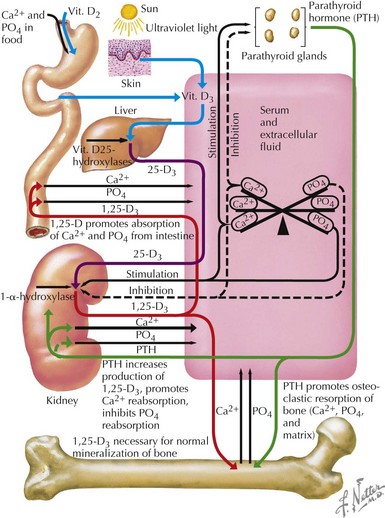
Most (99%) of the body’s calcium exists as hydroxyapatite in bone, with the remaining 1% present in extracellular fluids. Serum calcium exists in three fractions: 50% to 55% is free (ionized) calcium; about 10% is complexed with low-molecular-weight anions; and 35% to 40% is bound to proteins, mainly albumin and, to a lesser extent, globulins. The calciotropic hormones calcitriol (the fully active form of vitamin D) and parathyroid hormone (PTH) act on their target organs, kidney, intestines, and bone to regulate mineral homeostasis (Figure 69-1). Phosphatonins such as FGF23 also play important regulatory roles in mineral metabolism and complement the actions of other calciotropic hormones; phosphatonins decrease renal phosphorus reabsorption while reducing synthesis of calcitriol and secretion of PTH.


The principal source of vitamin D is the skin. High-energy ultraviolet B light penetrates the epidermis and cleaves 7-dehydrocholesterol to produce previtamin-D3. Previtamin D3 then undergoes a thermally induced isomerization to vitamin D3 (cholecalciferol) that takes 2 to 3 days to reach completion. Therefore, after a single sunlight exposure, cutaneous synthesis of vitamin D3 continues for many hours. It is not possible to generate too much vitamin D3 in the skin because prolonged sunlight exposure activates a mechanism that converts excess previtamin D3 and vitamin D3 to biologically inert products. Vitamin D can also be obtained from the diet, from plant sources as ergocalciferol (vitamin D2), and from animal sources as cholecalciferol (vitamin D3). Both of these forms of vitamin D are fat soluble and are absorbed from the small intestine into the lymphatics. About 50% of the vitamin D in chylomicrons is transferred to the plasma, where it circulates tightly bound to proteins, principally vitamin D–binding protein (DBP, also termed Gc protein).


Additional enzymatic steps are required to produce the fully active vitamin D metabolite calcitriol (also termed 1,25(OH)2D3). Dietary and endogenously produced vitamin D undergoes 25-hydroxylation in the liver by the cytochrome P450 enzyme CYP2R1 to form 25(OH)D. Subsequently, 25-(OH)D3 is directed to the kidney, where it is either converted to 24,25-dihydroxyvitamin D3 (an inactive derivative) or to 1,25-dihydroxyvitamin D3 (calcitriol). Activation to calcitriol requires hydroxylation by a 1α-hydroxylase enzyme (CYP 27B1) that is tightly regulated and is the rate-limiting step in the bioactivation of vitamin D: PTH increases production of calcitriol by stimulating CYP27B1 activity, and FGF23 decreases CYP27B1 activity.


PTH is synthesized as a pre-prohormone by parathyroid cells and processed to a mature 84-amino acid peptide (intact or whole PTH) that is stored in secretory granules. Extracellular ionized calcium is the principal regulator of PTH release and interacts with G protein–coupled calcium-sensing receptors that are expressed on the cell membrane. Low or decreasing concentrations of ionized calcium stimulate secretion of stored PTH within seconds and subsequently increase synthesis of new hormone. PTH acts directly on bone and kidney and indirectly on the intestine to increase the extracellular calcium concentration. After release into circulation, PTH has a half-life of only 6 to 8 minutes and is degraded rapidly to inactive (or less active) fragments by endopeptidases in the liver and kidney. PTH binds to receptors on the surface of target cells that are coupled via guanine–nucleotide binding (G) proteins to activation of adenylyl cyclase and phospholipase C, which increase intracellular concentrations of the second messengers cyclic AMP, inositol triphosphate, and calcium.


Acutely, PTH acts on bone to activate osteoclastic bone resorption, which releases calcium (and phosphorus) into the circulation within minutes. Chronically elevated levels of PTH increase the number of osteoblasts and osteoclasts and stimulate bone remodeling, which over time leads to decreased bone mass and osteoporosis.


In the kidney, PTH increases distal tubular reabsorption of calcium and decreases proximal tubular and thick ascending limb reabsorption of sodium, calcium, phosphate, and bicarbonate. PTH (and hypophosphatemia) stimulates renal 25(OH)D-1α-hydroxylase, which increases synthesis of 1,25(OH)2D3 and promotes intestinal absorption of calcium.


Whereas most extracellular phosphate is located in bone mineral in the form of hydroxyapatite, intracellular phosphate is in nucleotides and nucleic acids, phosphoproteins, and phospholipids. Therefore, phosphate’s important roles include maintenance of bone mineral, regulation of enzyme activity, and energy metabolism. Dietary phosphate is amply available and readily absorbed. Movement in and out of the bone mineral is regulated by PTH and 1,25(OH)2D3. In the kidney, PTH and phosphatonins (e.g., FGF23) inhibit phosphate transport by reducing membrane expression of Napi 2a and Napi 2c sodium-phosphate cotransporters in the proximal renal tubule cells.


Dynamics of Bone Homeostasis


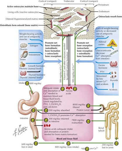
Skeletal development is a complex process sensitive to the hormonal, mechanical, and nutritional milieu of the bone. The shape and structure of bones are modified and renovated by two processes, modeling and remodeling. Remodeling is the major process in adults and does not result in a change of the bone shape. This process takes place in the basic bone multicellular units where bone resorption by osteoclasts is tightly coupled to bone formation by osteoblasts. Old or damaged bone is repaired by remodeling. By contrast, modeling occurs only during development and growth of the skeleton and facilitates new bone formation at a location different from the site of bone resorption. Growth in the diameter of the cortical shaft is the result of bone formation at the outer (periosteal) surface and bone resorption at the inner (endosteal) surface.


Bone modeling and remodeling are regulated by a variety of factors such as biomechanical loading, hormonal balance, acid–base status, and drug exposures (Figure 69-2). Bone adapts its strength in response to the magnitude and direction of the forces to which it is subjected. Mechanical forces on the skeleton arise primarily from muscle contraction. This capacity of bone to respond to mechanical loading with increased bone size and strength is greatest during growth, especially during puberty and adolescence. Increased production of estrogen and testosterone in addition to increased pulsatile secretion of growth hormone are the hormonal hallmarks of puberty. These events act in an anabolic manner on bone to promote net bone formation.


< div class='tao-gold-member'>

Stay updated, free articles. Join our Telegram channel

Jun 19, 2016 | Posted by in PEDIATRICS | Comments Off on Disorders of Calcium and Bone Metabolism

Full access? Get Clinical Tree

Get Clinical Tree app for offline access