Chapter 10 Diseases of the Cervix
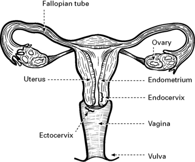
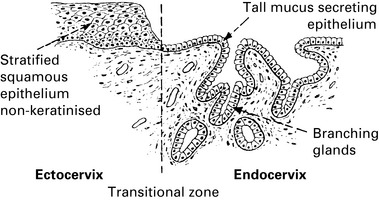
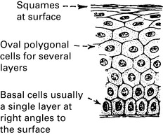
Normal cervical epithelium
Anatomy of the cervix
The cervix constitutes the lower third of the uterus.
It is in two parts, the endocervix and the ectocervix. Before puberty, the dividing line between the endo- and ectocervix is sharp, and is determined by the character of the lining epithelium: the ectocervix is covered with squamous epithelium and the endocervix is covered with columnar epithelium.
Benign squamous metaplasia
Stratification well defined with:
Under the influence of oestrogen (e.g. at puberty, during pregnancy or when the combined pill is taken), the cervix enlarges, so that the columnar epithelium of the endocervix can be seen on the ectocervix. This region, formerly the endocervix, which is now anatomically the ectocervix, is known as the ‘transformation zone’. The transformation zone is a dynamic area that responds to changes in endogenous or exogenous oestrogen. As it undergoes changes, it becomes a squamous metaplasia, which is a benign condition. The transformation zone is important as it is susceptible to infection with the human papilloma virus (HPV), which is known to lead to possible dysplastic change, and this in turn may lead to cervical cancer.
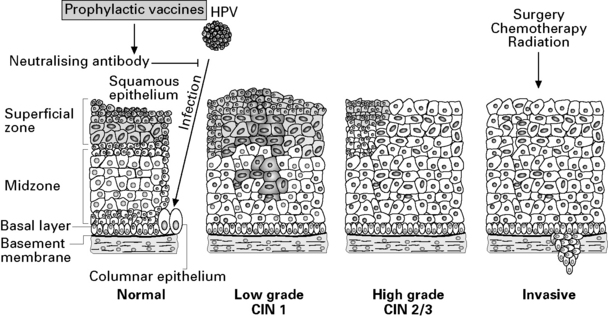
The role of human papilloma virus in cervical disease
Risk factors
Human Papilloma Virus (HPV) Infection
HPV infection is now known to be the viral agent required for the development of, initially, cervical intraepithelial neoplasia (CIN), and, ultimately, a cervical carcinoma. Approximately 80% of women will be infected with at least one of the more than 100 genotypes of HPV. 70–90% of women infected with HPV will clear the infection spontaneously within 1–3 years.
HPV is a DNA virus that can integrate into the human genome, and each genotype has a number. The most prevalent HPV genotypes in cervical carcinoma are 16 and 18. They are responsible for at least 70% of all cervical cancers.
The HPV genome encodes early (E) proteins that are responsible for viral regulation, cell transformation and late (L) proteins that make the viral capsule. HPV infects the basal cells and the expression of genes E6, E7 in high risk genotypes allows them to act as oncogenes via a variety of mechanisms to cause unregulated cell growth that may lead to dysplasia and cancer. Two tumour suppressor proteins that are affected by the E6/E7 gene products are p53 and retinoblastoma.
Suggested co-factors that may increase the likelihood of developing cervical cancer include: smoking, low socioeconomic status, immunosuppression (e.g. HIV infection, organ transplant), multiple sexual partners and combined oral contraceptive pill use for more than 10 years.
Screening for cervical cancer
Screening for cervical intraepithelial neoplasia (CIN) can be done by performing cervical smears and offering subsequent colposcopic assessment if significant dyskaryotic change is found. Treatment of CIN significantly reduces the incidence of and mortality associated with cervical cancer. For these reasons, many countries, including the UK, have set up a national screening programme.
Screening process
The Cervical Smear
A cervical smear is taken by a trained healthcare professional, often in their local practice. The cervix is visualised using a speculum. A wooden spatula or small plastic broom is placed in the cervical canal and rotated through 360°. The superficial layer of cells overlying the squamocolumnar junction is removed. If a wooden spatula is used, it is then smeared on a slide and fixed. If the broom is used, it is placed into a vial of liquid fixative (liquid-based cytology – LBC). Both methods then stain the specimen using Papanicolou’s method and the cells are examined under a microscope. The LBC technique is automated once the sample is sent for processing and this method produces cleaner specimens for analysis, thus reducing the number of unsatisfactory slides. If abnormal cells, dyskaryosis, are identified, the patients are referred for colposcopy.
In Scotland, the current cervical screening programme includes the following:
An abnormal smear is usually an indication for colposcopy (see p. 179).
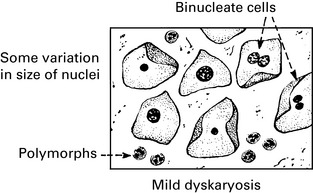
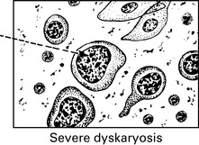
Cervical Smears
Interpretation of cervical smears
Smears are reported in the following categories:
The smear shows a mixture of normal cells and dyskaryotic (abnormal nuclei) cells.
The cells are almost all abnormal and show larger nuclei with coarse chromatin.
In general, the more severe the dyskaryosis, the more likely the patient is to have high grade CIN. However, even in women with a mildly dyskaryotic smear, the incidence of high grade CIN can be 50%.
A referral for colposcopy is made for all women with, moderate or severe dyskaryosis. A repeat smear is justified in women with inflammatory or borderline nuclear changes. However, if these abnormalities persist, colposcopy is indicated. Colposcopy is also indicated if there are three or more abnormal smear tests of any grade in a 10-year interval.
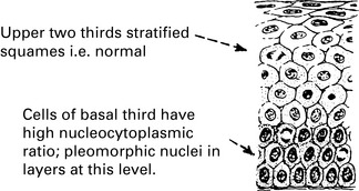
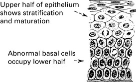
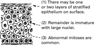
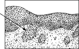
Premalignant and malignant cervical disease
Dysplasia
Dysplasia occurs when the epithelium shows changes such as nuclear enlargement, increased nucleocytoplasmic ratio and abnormal mitoses. It is regarded as the first step in a series of changes that may lead to cervical intraepithelial neoplasia (CIN) and subsequently to invasive carcinoma. The pathological features of mild, moderate and severe dysplasia are shown. In practice there can be inter- and intra-observer variation between these categories.
Microinvasive cervical carcinoma
The histological appearance of an early cervical invasion with spread through the basement membrane is shown here.
Risk of progression of cervical intraepithelial neoplasia
CIN has the potential to develop into a malignant cervical disease but it may also regress or persist as CIN without undergoing any malignant change. The risk of developing cervical cancer following dysplasia is 10–20%. The risk is also influenced by the grade of CIN, oncogenic potential of the HPV type, and environmental factors such as smoking and the immune status of a patient.

Stay updated, free articles. Join our Telegram channel

Full access? Get Clinical Tree

