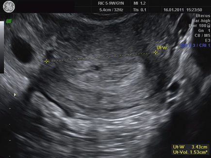
Fig. 2.1
Longitudinal view demonstrating regular thickened endometrium

Fig. 2.2
Transverse view demonstrating intrauterine pseudosac and isthmic tubal pregnancy to the left of the uterus. (These two ultrasound pictures (Figs. 2.1 and 2.2) were taken from the same patient. Longitudinal view demonstrates a normally appearing luteal phase endometrium while the transverse view reveals a pseudosac and a clearly visible ectopic pregnancy in the isthmic portion of the fallopian tube. With beta-hCG levels of 1500 IU/mL this ectopic pregnancy could easily have been missed. (Courtesy Arnon Agmon, MD)
References
1.
2.
3.
4.
Kirk E, Papageorghiou AT, Condous G, Tan L, Bora S, Bourne T. The diagnostic effectiveness of an initial transvaginal scan in detecting ectopic pregnancy. Hum Reprod. 2007;22:2824–8.CrossRefPubMed
Stay updated, free articles. Join our Telegram channel

Full access? Get Clinical Tree


