Chapter 44 Difficulty Breathing (Case 14)
Patient Care
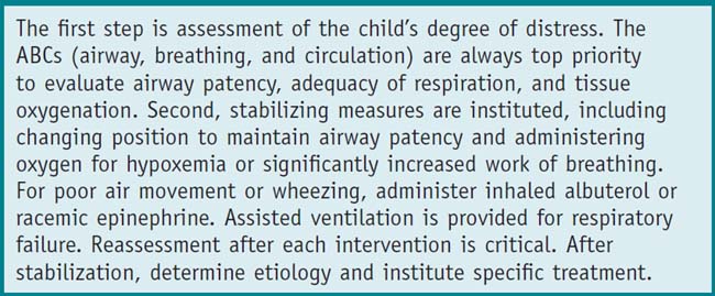
Clinical Thinking
• What is my first impression? A child who is pink, breathing, and mildly hypoxic but talking is mildly distressed. A child who has rapid shallow breathing, deep retractions, or appears tired is severely distressed.
• What is the degree of activity and interaction? A well toddler will generally resist being examined, but if in significant distress may offer no objection.
• Is there stridor to suggest extrathoracic obstruction, such as in croup, or expiratory wheezing to suggest intrathoracic obstruction?
History
• Any prior episodes of difficulty breathing? Any hospitalizations, prior intubation, or intensive care admissions?
Physical Examination
• Check vital signs: Elevated temperature for infection, respiratory rate and pulse oximetry to assess oxygenation, heart rate and blood pressure for perfusion.
• Focus on the respiratory examination: Note respiratory effort, adequacy of ventilation, any asymmetry, nasal flaring, and presence of retractions (subcostal, intercostal, or supraclavicular). Check for drooling and note child’s position of comfort (e.g., sitting up, tripod position).
• Biphasic stridor indicates obstruction at the level of the distal larynx/trachea. Upper airway rhonchi, suggesting nasal or postnasal congestion, may also be biphasic.
• Dullness to percussion suggests an effusion or mass, although these findings are often difficult to localize in infants and toddlers.
Tests for Consideration
Clinical Entities: Medical Knowledge
| Croup | |
|---|---|
| Pϕ | Croup, or laryngotracheobronchitis, is a viral infection causing edema and inflammation in the subglottis, most commonly resulting from parainfluenza. Other causes include adenovirus, respiratory syncytial virus (RSV), human metapneumovirus (HMPV), and influenza. Spasmodic (noninfectious, typically allergic) croup presents acutely, usually in the middle of the night without a viral prodrome or fever. It tends to recur, and the etiology is unclear. |
| TP | Children with croup are typically 6 months to 3 years of age and present with acute onset of inspiratory stridor, hoarseness and barky cough a few days after onset of upper respiratory symptoms. Symptoms are worse at night and with agitation. There may be high fever and significant respiratory distress with supraclavicular retractions, tachypnea, and hypoxemia. |
| Dx | The classic triad is inspiratory stridor, barky cough, and hoarseness. Clinical severity is determined using a croup severity score, which assesses inspiratory stridor, retractions, air entry, cyanosis, and level of consciousness. Although not typically indicated, a posteroanterior neck radiograph may demonstrate subglottic narrowing (steeple sign). |
| Tx | Humidity, cool night air, nebulized racemic epinephrine, corticosteroids (a single dose of dexamethasone [0.6 mg/kg IM or PO]) and supplemental oxygen are administered as needed. The need for hospitalization is assessed based on severity of presentation, response to acute treatment, and availability of follow-up. See Nelson Essentials 107. |
| Bronchiolitis | |
|---|---|
| Pϕ | In bronchiolitis, a virus infects distal bronchiolar epithelium, causing inflammation, bronchiolar cell necrosis, ciliary disruption, airway edema, mucus production, and sloughed epithelial cells, which all contribute to airway obstruction and atelectasis. The resulting ventilation-perfusion mismatch leads to hypoxemia with increased work of breathing. RSV is the most common cause. Others include HMPV, adenovirus, influenza, parainfluenza, rhinovirus, and human bocavirus. |
| TP | Occurring up to 2 years of age, the initial symptoms are nasal congestion, rhinorrhea, low-grade fever, and decreased feeding. With lower airway involvement there is wheezing and a tight cough, tachypnea, accessory muscle use with retractions, nasal flaring, and/or grunting. In more severe cases there may be hypoxemia or apnea. More severe cases occur in children under 6 months. Distinguishing viral-induced wheezing from an asthma exacerbation can be difficult. With increased work of breathing and hypoxemia, infants may have difficulty maintaining adequate oral intake and risk dehydration. |
| Dx | Diagnosis is made clinically and includes assessing illness severity. Specific viral testing may be helpful for admission cohorting. Chest radiograph may show hyperinflation with peribronchial thickening and subsegmental atelectasis. Focal densities occur in severe cases. Risk factors for severe disease include prematurity, age less than 12 weeks, congenital heart disease, neurologic disorders, immunodeficiencies, and pulmonary disease. |
| Tx | Supportive care is the mainstay of therapy. Consider supplemental oxygen, inhaled albuterol or racemic epinephrine, and continue if improvement is noted. Small feeding volumes may reduce gastric distention. Respiratory status, age, and duration of illness will determine need for hospitalization. With outpatient management, young infants and those early in the course need frequent office visits to monitor hydration status and work of breathing. See Nelson Essentials 109. |
| Pneumonia | |
|---|---|
| Pϕ | Pneumonia is a lower tract infection involving the bronchioles, alveoli, and/or interstitium, and the most common bacterial causes are Streptococcus pneumoniae and increasingly Staphylococcus aureus. Viruses and atypical bacteria (Mycoplasma pneumoniae) infect airway epithelial cells, producing a diffuse interstitial pattern. Pneumonia may occur via several mechanisms: as a primary infection of the lower tract, in association with an upper tract infection (common cold), from aspiration, or via hematogenous spread. Parapneumonic effusions and empyema are more common with streptococcal and staphylococcal infections, particularly community-acquired methicillin-resistant S. aureus (CA-MRSA). Likely pathogens vary based on age, environment, and risk factors (see Chapter 31, Cough, and Chapter 46, Neonatal Fever). |
| TP | A child with bacterial pneumonia typically presents with fever, tachypnea, and cough, and often malaise, headache, and abdominal pain. On examination focal crackles and/or wheezing may be heard. Interstitial pneumonia presents with fever, cough, and diffuse lung findings, though differentiation of “typical” from “atypical” pneumonia may be difficult. Young infants may have fever and tachypnea without localizing lung findings. |
| Dx | Diagnosis can be made clinically in a nontoxic child with classic physical findings. Focal consolidation on radiograph often reflects bacterial pneumonia. Aspiration more commonly occurs in the right upper lobe. Dullness to percussion suggests an effusion. Decubitus chest radiograph, ultrasound, or CT scan further delineates pleural fluid. Viral pathogens and atypical bacteria usually produce a diffuse interstitial pattern. |
| Tx | Antibiotics that cover gram-positive organisms should be started. High-dose amoxicillin/ampicillin targets intermediately resistant pneumococcus. Macrolides may be used in penicillin-allergic patients and in suspected M. pneumoniae. Antibiotics are not indicated for viral processes. Persistently febrile and ill-appearing children should have a repeat chest radiograph to exclude parapneumonic effusion or empyema. With sizeable collections, drainage with video-assisted thoracoscopy (VATS) or chest tube placement may be needed. Cell count, Gram stain, and culture guide antibiotic treatment. Aspiration pneumonia requires coverage of gram-negative and perhaps anaerobic organisms. MRSA is usually treated with clindamycin, vancomycin, or linezolid, depending on illness severity and regional sensitivity patterns. See Nelson Essentials 110. |
| Asthma | |
|---|---|
| Pϕ | Asthma is characterized by recurrent reversible airflow obstruction, inflammation, and hyperresponsiveness of the lower airways. Common triggers are viral respiratory infections, environmental tobacco smoke, and allergens such as dust mites, animal dander, molds, and pollen. Airway edema with mucus plugging causes air trapping with hyperinflation and atelectasis, resulting in ventilation–perfusion mismatch. |
| TP | Presentation varies, but typical findings are polyphonic bilateral expiratory wheezing and cough, although wheezing may be inspiratory or absent with severely compromised airflow. Wheezing is absent in cough-variant asthma. |
| Dx | Diagnosis is made clinically, based on symptoms and physical examination findings, along with response to bronchodilator therapy. Baseline peak expiratory flow (PEF) is decreased during an exacerbation, as is forced expiratory volume (FEV1) on spirometry. There may be atopic findings such as eczema and seasonal allergies. There is overlap in presentation between asthma and bronchiolitis in children under 2 years. A prior history of wheezing and a family history of asthma or atopy can help solidify the diagnosis. |
| Tx | First-line treatment, after assessing the ABCs and providing supplemental oxygen, is to reverse bronchoconstriction. Albuterol, a short-acting beta agonist, is most commonly used, either by nebulization or inhaler. An aerosolized anticholinergic such as ipratropium may be useful on initial presentation. Additional medications include terbutaline, epinephrine, and intravenous magnesium sulfate. Inflammation is treated with systemic corticosteroids. Consider a chest radiograph if concern for complications: pneumonia, pneumothorax, pneumomediastinum, or significant atelectasis. All patients should be discharged with an asthma action plan and close follow-up (see Chapter 31, Cough, and Chapter 87, Status Asthmaticus). See Nelson Essentials 78. |
| Foreign Body | |
|---|---|
| Pϕ | Most foreign bodies are lodged in the bronchi, most commonly in the right lung. A laryngotracheal FB causes significantly higher morbidity and mortality than bronchial aspirations. Common aspirates include seeds, nuts (especially peanuts), and small toys. |
| TP | Respiratory distress or altered mental status require immediate intervention with bronchoscopy and appropriate respiratory support to relieve airway obstruction. Cyanosis, hoarseness, stridor, and acute respiratory distress suggest laryngotracheal aspiration. Nonemergent presentations are more common: following the acute event, after coughing and gagging have subsided, the child may be asymptomatic; thus an accurate history is key in making the diagnosis. This is especially true with a bronchial FB, in which the classic triad of wheeze, cough, and diminished breath sounds occurs in only 50% of cases. Cough or wheeze that fails to resolve should raise concern for an aspirated FB. |
| Dx | Radiopaque objects such as coins may be seen on the radiograph, though most objects are radiolucent. With a bronchial FB, radiographic findings include hyperinflation, atelectasis, mediastinal shift, and pneumonia. A lateral decubitus chest film may demonstrate persistent air trapping in the dependent lung. |
| Tx | Bronchoscopy is the treatment of choice. After removal of the FB, the airway is evaluated for smaller fragments and general inflammation. Gram stain and culture of fluid are performed to excludea secondary pneumonia. Antibiotics should be started accordingly. See Nelson Essentials 136. |
Only gold members can continue reading. Log In or Register to continue
Stay updated, free articles. Join our Telegram channel

Full access? Get Clinical Tree


