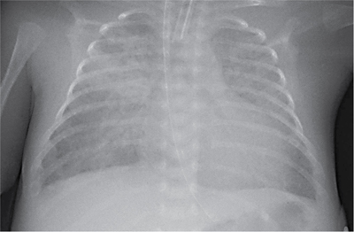
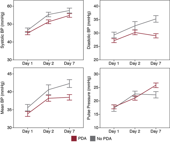
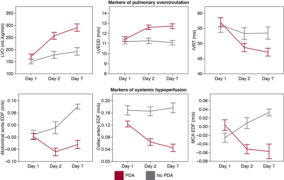
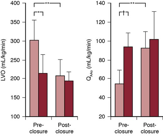
Audrey Hébert, Afif Faisal El-Khuffash, Shahab Noori, Patrick J. McNamara Key points The diagnosis and management of a patent ductus arteriosus (PDA) with cardiorespiratory, and thus clinical, relevance in preterm neonates poses a major challenge in neonatal medicine. It is the most common cardiovascular abnormality in premature infants. Most (∼70%) infants born at a gestational age (GA) of less than 29 weeks will have a persistent PDA by the end of the first postnatal week.1 PDA is associated with several morbidities and mortality; however, a cause-and-effect relationship between the presence of a PDA and important short- and long-term clinical outcomes has not been definitively established. In addition, there are limitations due to study design of randomized control trials and the retrospective nature of cohort studies reporting clinical outcomes and the impact of various approaches to treatment (conservative, medical, device and surgical). More than 50% have a high rate of open-label treatment in the control arm and high treatment failure rate in the intervention arm.2 There is considerable heterogeneity in infants included in PDA trials due to lack of a standardized definition of hemodynamic significance, resulting in inclusion of patients who may not benefit from PDA treatment.3 As a result, the impact of PDA treatment on outcomes and treatment strategies (particularly modality and timing) varies between centers.4 Finally, there continues to be a lack of physician equipoise such that the most vulnerable infants, potentially at greatest risk of PDA-attributable morbidity, are not always enrolled in clinical trials.5 The ductus arteriosus (DA) connects the main pulmonary artery to the descending aorta and is necessary for fetal survival. In the fetus the left ventricle (LV) receives oxygenated blood, returning from the placenta via the inferior vena cava and through the foramen ovale, and delivers it mainly to the upper part of the body. The right ventricle (RV) receives most blood draining from the superior vena cava (SVC) and a proportionately lower amount of oxygenated blood from the umbilical venous system. Due to high pulmonary vascular resistance (PVR), most (80% to 90% depending on the gestational age of the fetus) of right ventricular output flows from the pulmonary artery to the descending aorta across the DA; hence the DA modulates flow to the lower part of the body. Similarly, the patent foramen ovale (PFO) is the route that modulates the delivery of oxygenated blood to the head and neck. After birth, LV afterload rises suddenly due to loss of the low resistance placental circulation. This is accompanied by lung aeration, which promotes a fall in PVR and an increase in pulmonary blood flow. This results in a change in the organ of gas exchange from the placenta to the lungs. The DA eventually closes (first functionally and then anatomically) over the subsequent days in term infants. However, in preterm infants the DA can remain patent for a prolonged period of time for a variety of reasons. In term infants closure of the PDA occurs within the first 48 hours after birth. Closure of the DA occurs in two phases. The first phase (within the first hours after birth), termed “functional closure,” involves narrowing of the lumen by smooth muscle constriction. The second phase, termed “anatomic remodeling”, consists of occlusion of the residual lumen by extensive neointimal thickening and loss of muscle media smooth muscle over the next few days. The rate and degree of initial “functional” closure are determined by the balance between factors (mediators, second messengers and channels, among others) that favor constriction (oxygen, endothelin, calcium channels, catecholamines, and Rho kinase) and those that oppose it (intraluminal pressure, prostaglandins [PGs], nitric oxide [NO], carbon monoxide, potassium channels, cyclic adenosine monophosphate [AMP], and cyclic guanosine monophosphate). PGs play a key role in the regulation of ductal tone, especially during the first few postnatal weeks. Of these, PGE2 is the most important factor in the regulation of DA tone during fetal development and acts on G protein–coupled E-prostanoid receptors to maintain ductal patency. It is generated from arachidonic acid by cyclooxygenase-1 (COX-1) and COX-2, the COX component of PG-H2 synthase, followed by peroxidation by the same enzyme complex and, finally, by the action of PGE synthase (see Chapter 5). COX-2 plays a major role in maintaining ductal patency during fetal life.6 The current approach to medical therapy exploits this mechanism by the use of nonselective COX inhibitors (such as indomethacin and ibuprofen) and also by the use of acetaminophen, a peroxidase inhibitor, to close the DA postnatally. Low oxygen tension in the fetus is another important factor for maintaining ductal patency.7 Following birth, the rise in oxygen tension promotes an oxygen-mediated constriction that is facilitated by the inhibition of the potassium voltage channels (KV channels) present on the ductal smooth muscle cells and function to keep the cells in a hyperpolarized state.8 The presence of oxygen leads to depolarization, which in turn activates L-type calcium channels, allowing an influx of calcium into the smooth muscle cells causing constriction. A counter-mechanism via the mitochondrial electron transport chain serves as the intrinsic oxygen-sensing mechanism that regulates this constrictive effect via formation of reactive oxygen radicals, which inhibit KV channels.9 Interestingly, in vitro studies using rings of human DA tissue incubated in relatively low oxygen tension conditions (to mimic conditions of prematurity) for several days selectively fail to constrict in response to oxygen. This may explain, at least in part, failure of the DA to close in preterm infants. The fall in PG levels following birth (due to the loss of placental PG production and increase in its removal by the lungs), accompanied by the rise in oxygen tension, promotes functional closure of the DA over the first 24–48 postnatal hours. After functional DA closure is achieved, the smooth muscle cells migrate from the media to the subendothelial layer, leading to neointimal formation.10 Expansion of the neointima forms protrusions, or mounds, that permanently occlude the already constricted lumen.11,12 This process results in an interruption of the blood supply to the innermost cellular layer, resulting in hypoxia and cell death.13 The presence of intramural vasa vasorum is essential to ensure adequate provision of oxygen and nutrition to the thicker wall of the DA at term. During postnatal constriction, the intramural tissue pressure obliterates vasa vasorum flow in the muscle media. The ensuing ischemic and hypoxic insult inhibits local PGE2 and NO production, induces local production of hypoxia-inducible factors (HIF) like HIF-1α and vascular endothelial growth factor (VEGF), and produces smooth muscle apoptosis in the muscle media. In addition, monocytes/macrophages adhere to the ductus wall and appear to be necessary for ductus remodeling.14 In contrast, in preterm infants the DA frequently fails to constrict or undergo anatomic remodeling after birth. In fact, a study by Semberova et al showed that the median time to PDA closure was 71 days in infants <26 weeks’ gestation.15 There is, however, little information regarding the biological mechanisms that contribute to late spontaneous closure. The incidence of persistent PDA is inversely related to gestational age due to several mechanisms.16 The intrinsic tone of the extremely immature ductus (<70% of gestation) is decreased compared with the ductus at term.17 This may be due to the presence of immature smooth muscle myosin isoforms, with a weaker contractile capacity,18–21 and to decreased Rho kinase expression and activity.22–24 Calcium entry through L-type calcium channels also appears to be impaired in the immature ductus.23–25 In addition, the potassium channels, which inhibit ductus contraction, change during gestation from KCa channels not regulated by oxygen tension to KV channels, which can be inhibited by increased oxygen concentrations.26–28 The reduced expression and function of the putative oxygen-sensing KV channels in the immature ductus appear to contribute to ductus patency in several animal species.24,26,28 In most mammalian species the major factor that prevents the preterm ductus from constricting after birth is its increased sensitivity to the vasodilating effects of PGE2 and NO.29 The increased sensitivity of the preterm ductus to PGE2 is due to increased cyclic AMP signaling. There is both increased cyclic AMP production, due to enhanced PG receptor coupling with adenylyl cyclase, and decreased cyclic AMP degradation by phosphodiesterase in the preterm ductus.30,31 As a result, inhibitors of PG production (e.g., indomethacin, ibuprofen, mefenamic acid, paracetamol) are usually effective agents in promoting ductus closure in the premature infant. Premature infants also have elevated circulating concentrations of PGE2 due to the decreased ability of the premature lung to clear circulating PGE2.32 In the preterm newborn circulating concentrations of PGE2 can reach the pharmacologic range during episodes of bacteremia and necrotizing enterocolitis and are often associated with reopening of a previously constricted DA.33 Little is known about the factors responsible for the changes that occur with advanced gestation. A recent study showed gene expression in pathways involved with oxygen-induced constriction, contractile protein maturation, tissue remodeling, and PG and NO signaling alter according to advancing GA.34 Prenatal administration of glucocorticoids reduces the incidence of PDA in premature humans and animals.35–40 Although postnatal glucocorticoid or corticosteroid administration also reduces the incidence of PDA, it is not clear whether this is a direct effect on ductal biology or an indirect effect on ambient conditions which promote ongoing ductal patency. In addition, it is important to recognize that glucocorticoid (dexamethasone) or corticosteroid (hydrocortisone) treatment, especially if it is given in the immediate postnatal period or combined with administration of COX inhibitors, respectively, has been associated with increased incidence of several other neonatal morbidities.41,42 The patient’s genetic background also seems to play a significant role in determining persistent ductus patency. Several single nucleotide polymorphisms in candidate genes have been identified that are associated with PDA in preterm infants: angiotensin receptor (ATR) type 1,43 interferon-gamma (IFN-γ),44 estrogen receptor-alpha PvuII,45 transcription factor AP-2B (TFAP2B), PGI synthase, and TRAF1.46 Studies suggest that an interaction between preterm birth and TFAP2B may be responsible for the PDAs that occur in some preterm infants: TFAP2B is uniquely expressed in ductus smooth muscle and regulates other genes that are important in ductus smooth muscle development.47 Mutations in TFAP2B result in patency of the DA in mice and humans48 and TFAP2B polymorphisms are associated with the PDA in preterm infants (especially those that are unresponsive to indomethacin).49 Expression of SLCO2A1 and NOS3 genes (involved with PG reuptake/metabolism and NO production, respectively) is decreased in the DA from non-Caucasians.34 This may lead to an increase in PG and decrease in NO concentrations, thereby making ductal patency more PG dependent and possibly explaining the clinical finding of a better response to indomethacin in non-Caucasians.34,49,50 Recently, an association was shown between two single nucleotide polymorphisms in CYP2C9, rs2153628, and rs1799853, and indomethacin response for the treatment of PDA. Findings suggest that response to indomethacin in the closure of PDA may be influenced by polymorphisms associated with altered indomethacin metabolism.51 There is, however, little data on the genetic determinants of acetaminophen response. Neointimal mounds are less well developed and often fail to occlude the lumen in preterm infants (especially those born before 28 weeks’ gestation). The preterm ductus is a much thinner vessel than the full-term ductus; therefore there is no need for vasa vasorum because the vessel wall is nourished with oxygen via diffusion through luminal blood flow (vasa vasorum first appear in the outer ductus wall after 28 weeks’ gestation). As a result, unless the ductus lumen is completely obliterated, the preterm ductus is less likely to develop profound hypoxia as it constricts after birth. Without a strong hypoxic signal, neointimal expansion is markedly diminished, resulting in mounds that fail to occlude the residual lumen11,14,52,53 During fetal life, low systemic vascular resistance (SVR) due to the low resistance placenta, combined with elevated PVR, results in pulmonary artery–to-aorta (“right-to-left”) flow across the DA. During normal neonatal transition, increased SVR associated with umbilical cord clamping occurs in parallel to a longitudinal decrease in PVR precipitated by ventilation of the lungs and an increase in pulmonary blood flow. The degree of right-to-left ductal shunt is approximately 50% within 5 minutes of birth, becoming mostly left to right by 10 to 20 minutes, and is entirely left to right by 24 hours of age in most healthy neonates.54,55 In preterm neonates the size and direction of the ductal shunt will have a variable impact on pulmonary and systemic hemodynamics. The role of the PDA shunt may be conceptualized within a physiologic continuum that extends from a life-sustaining conduit, neutral bystander, to a pathologic entity. In infants with critical congenital heart disease patency of the DA may be necessary to support pulmonary (e.g., tricuspid atresia) or systemic (e.g., critical aortic stenosis) blood flow. In acute pulmonary hypertension (aPH) of the newborn postnatal failure of pulmonary arterioles to relax (e.g., due to asphyxia, respiratory distress syndrome) results in high PVR and persistence of a right-to-left ductal shunt. The latter shunt may reduce right ventricular afterload and support post-ductal systemic blood flow, albeit with deoxygenated blood. PDA closure in this setting will negatively impact RV function and the adequacy of systemic blood flow. A bidirectional shunt in milder cases of aPH may play a neutral role, merely permitting the noninvasive estimation of the systemic-pulmonary pressure gradient. If the DA remains patent after birth, preterm infants who experience the expected fall in PVR may be susceptible to the effects of a large systemic-to-pulmonary (left-to-right) shunt. Blood flows across the PDA continuously in systole and diastole, resulting in volume overload of the pulmonary artery, pulmonary veins, and left heart. Shunt volume (Q) is directly proportional to the fourth power of the ductal radius (r) and the aortopulmonary pressure gradient and is inversely proportional to the ductal length (L) and blood viscosity (n). It is important to consider the relative contributions of each component to shunt volume. Increased pulmonary blood flow (termed pulmonary overcirculation) may lead to alveolar edema, reduced pulmonary compliance, and increased need for respiratory support.56 Increased blood flow to the left heart results in dilatation and increased end-diastolic pressures in the left ventricle and atrium. In preterm infants with intrinsic immaturity of LV compliance and diastolic function, the increase in end-diastolic pressure may contribute to the evolution of pulmonary venous hypertension and pulmonary hemorrhage. The increase in pulmonary blood flow, due to a large left-to-right shunt, occurs at the expense of systemic blood flow (referred to as ductal steal), which may result in end-organ hypoperfusion and consequential morbidities (e.g. necrotizing enterocolitis, acute tubular necrosis).57 In addition, ductal steal from the descending aorta, shorter diastolic and coronary perfusion times due to tachycardia, and increased myocardial oxygen demand may result in subendocardial ischemia. This pathophysiologic cascade is thought to explain, at least in part, the relation between a PDA and adverse outcomes. Cardiac output is the result of the interactions between preload, afterload, intrinsic myocardial contractility, and heart rate. Under normal conditions, and in the absence of a PDA, LV output (LVO) in a neonate is in the range of 150–300 mL/min/kg. The presence of a PDA results in increased pulmonary blood flow and both left atrial (LA) and LV volume loading. In a prospective observational study, using two-dimensional speckle tracking echocardiography, most infants with a PDA displayed signs of LA dysfunction due to increased volume load. PDA diameter was found to be an independent contributor to poor LA contraction.58 Studies have consistently shown a higher LV end-diastolic volume (preload) when the DA is open with a predominantly left-to-right shunting pattern. According to the Starling curve, the increase in myocardial muscle fiber stretch from higher preload augments stroke volume. Indeed, most studies have demonstrated increased LVO in the presence of a PDA with a predominant left-to-right shunt.59–68 In the presence of a PDA the low-resistance pulmonary vascular bed is in parallel with the systemic vascular bed. This results in a reduction of LV afterload, which, in combination with the increased preload, enhances the myocardium’s ability to increase its stroke volume. Traditionally, the presence of a PFO was thought to alter the effects of a PDA on LV stroke volume by exclusively decompressing the left atrium.69 Interestingly, a recent study has shown that larger atrial communication in the first week of life may be a surrogate marker of hemodynamically significant PDA rather than shunt volume modulator. The presence of a large atrial communication may in fact increase the risk of ventilator requirement and composite outcome of death or CLD.70 There are important differences in both the structure and function of the myocardium between preterm and term neonates, and older children and adults. These differences place the immature myocardium at a disadvantage as far as contractility is concerned.71 Furthermore, because coronary blood flow takes place primarily during diastole, myocardial performance might be adversely affected if diastolic blood pressure is low in the presence of a high-volume PDA shunt. Previous studies have suggested that myocardial ischemia may occur in the presence of a hemodynamically significant PDA (hsPDA).72 More recently, studies have demonstrated compromised coronary artery perfusion and the presence of high cardiac-specific troponin levels (indicative of myocardial damage) in the presence of a PDA, suggesting a detrimental effect on myocardial perfusion and potential ischemia.73,74 As premature infants have a less compliant myocardium than term infants, ventricular filling becomes dependent on the late diastolic phase of atrial contraction. In the setting of higher LV preload impaired diastolic function can lead to an increase in LA pressure and secondary pulmonary venous hypertension, potentially creating the biologic milieu that increases the risk of hemorrhagic pulmonary edema. Some authors have suggested that because higher preload is associated with a greater stretch of myocardial fibers75; therefore myocardial contractility should increase in the presence of a PDA concurrent with the increased LVO. On the contrary, the lack of change in myocardial contractility, in the presence of a PDA, could also suggest a relative deterioration of myocardial function based on increased demands. However, using a relatively load-independent measure of myocardial contractility, Barlow et al. showed that hsPDA had no effect on contractility.76 More recent studies, using more advanced functional parameters such as strain analyses, have also failed to demonstrate worsening function in the presence of the PDA.77–79 Preservation of LV function occurs despite major changes in LV morphology over the first 4 postnatal weeks. This includes an increase in LA volume, LV end-diastolic volume, sphericity index (indicating a more globular heart), and filling pressure.80 The potential impact of a PDA on RV function remains poorly understood. Changes seen in the LV are typically a consequence of pulmonary overcirculation, as described previously. Conversely, systemic hypoperfusion may result in a reduction in RV preload even in the presence of a left-to-right PFO shunt. In addition, prolonged exposure to increased pulmonary blood flow may promote an increase in PVR and a resultant increase in RV afterload. Recent studies have demonstrated reduced RV function as early as day 7 in infants with a large PDA.81 The clinical relevance of these changes to heart function and morphology and their potential impact on the evolution of PDA-associated morbidities is currently unknown. Blood pressure (BP) is the product of the interaction between cardiac output and peripheral vascular resistance (see Chapter 3). In general, systolic BP is primarily affected by changes in stroke volume, whereas diastolic BP is mainly reflective of changes in peripheral vascular resistance. Traditionally, low diastolic BP has been considered the hallmark of an hsPDA, and many studies have supported this notion.66,77 Studies that specifically looked at the relationship between BP and PDA have shown similar decreases in both systolic and diastolic BP (and therefore no change in the pulse pressure), at least during the first postnatal week.82,83 However, differences in BP and pulse pressure may be influenced by the location of BP measurements, pre- vs post-ductal. Typically, BP is measured from the umbilical arterial catheter (thus post-ductal) in the first postnatal week. Pre-ductal systolic BP is likely to be reflective of the increase in LV preload and higher than post-ductal values in patients with high-volume run-off through the PDA. This may generate discordance in systolic BP measurements, although this physiological hypothesis has yet to be confirmed in clinical trials. Older infants born at weights between 1000 and 1500 g with a PDA have slight, but clinically nonsignificant, decreases in systolic, diastolic, and mean BP. In contrast, infants born at less than 1000 g and with a PDA have both clinically and statistically lower systolic, diastolic, and mean BP but no change in pulse pressure.83 Because stroke volume increases and vascular resistance decreases in the presence of a PDA, one might expect that systolic BP would be maintained despite the decrease in diastolic pressure. As mentioned previously, the recorded systolic BP in most studies to date is post-ductal, which is not likely to reflect shunt-driven increases in LVO. In addition, cardiac output, ductal shunt volume, and peripheral resistance were not measured in any of these studies, making it difficult to accurately characterize the pulse pressure changes in the pre- vs post-ductal circulation. Therefore the lack of a discordance in pulse pressure from BP measured through an umbilical arterial line (post-ductal) could be used to exclude the presence of a high-volume PDA shunt. In immature animals a decrease in the diastolic and mean BP occurs even when the shunt is small, whereas a significant decrease in systolic BP occurs only when the PDA shunt is moderate or large.61 In a more recent cohort of 141 preterm infants, born less than 29 weeks’ gestation, systolic BP in infants with a PDA by the first postnatal week was only slightly lower than those without a PDA. It is not clear, however, if the location of BP measurement (pre- vs post-ductal) was consistent. On the contrary, diastolic and mean blood pressure was lower by the end of the first postnatal week, which translates into a higher pulse pressure (Figure 16.1).1 In this group LVO was higher and diastolic flow in systemic vessels was lower, possibly explaining those findings (Figure 16.2). PDA may also contribute to the development of hypotension, even during the transitional period, in patients with rapid drop in pulmonary vascular resistance. A study found evidence for a possible role of a moderate-large PDA in vasopressor-dependent hypotension.84 Similarly, PDA is reported to be an independent risk factor for refractory hypotension.85 Despite the ability of the LV to increase its output in the face of a left-to-right PDA shunt, organ blood flow distribution is significantly altered. Interestingly, redistribution of systemic blood flow occurs even with small shunts.61 Blood flow to the skin, bone, and skeletal muscle is most likely to be affected first by the left-to-right shunt. The organs affected thereafter are the gastrointestinal tract and kidneys, due to a combination of decreased perfusion pressure (ductal steal) and localized vasoconstriction (compensatory measure). Indeed, mesenteric blood flow is decreased in both fasting and fed states in the presence of a PDA.86 Clinically important decreases in blood flow to these organs may occur before there are signs of LV compromise.65,66 In addition, treatment strategies used to facilitate closure of the PDA, such as indomethacin, may have an effect on organ blood flow independent of the hemodynamic changes associated with the presence of an hsPDA.87 Data from a large national database, however, showed that the risk of necrotizing enterocolitis (NEC) was lower in patients with a PDA who received indomethacin vs those who remained untreated.88 Although cerebral blood flow (CBF) has also been assessed by near-infrared spectroscopy (NIRS) and magnetic resonance imaging (MRI) (see later and Chapter 13), blood flow velocity, measured by the Doppler technique, has been the most frequently used technique to assess changes in organ blood flow in the human neonate. In animal experimental models organ blood flow has also been measured by the microsphere technique or direct flow measurements. As discussed in Chapter 12 in detail, each of these techniques has significant limitations. Unfortunately, it is not currently feasible to continuously measure absolute blood flow to different organs in human neonates. Using the Doppler technique with ultrasonography, the amount of blood flowing through a vessel is a function of both the vessel diameter and mean blood flow velocity. Because of the small size of the neonatal blood vessels (e.g., anterior cerebral artery [ACA] or middle cerebral artery [MCA]), accurate measurement of vessel diameter is not possible. In addition, the Doppler technique assumes that the diameter of the vessel remains constant during the cardiac cycle, a notion that has been repeatedly challenged. Despite these limitations, Doppler velocity measurements and velocity-derived indices have been shown to have acceptable correlations with more invasive measures of organ blood flow.89–91 The most commonly used Doppler indicators of organ blood flow are systolic, diastolic, and mean blood flow velocities; velocity time integral; pulsatility index (PI); and resistive index (RI). Because the PI and RI are inversely related to flow, and directly related to vascular resistance, an increase in the PI or RI indicates a reduction in organ blood flow and/or an increase in the vascular resistance of the organ. Although some studies suggest that CBF is maintained in the presence of an hsPDA,60,66 most studies have shown a decrease in flow and a disturbance in cerebral hemodynamics.61 Furthermore, indomethacin, one of the drugs used for pharmacologic closure of the PDA, has a direct, albeit transient, vasoconstrictive effect on the cerebral circulation, which is likely independent of the drug’s effect on the COX enzyme.92,93 Using the Doppler technique, Perlman et al. demonstrated a decrease in diastolic blood flow velocity in the ACA of preterm infants in the presence of hsPDA.94 Similarly, Lemmers et al. reported that an hsPDA had a negative impact on cerebral oxygenation that resolved after treatment with indomethacin.95 Investigators have also observed retrograde diastolic flow and increased PI in the ACA in the presence of a PDA.96 In contrast, Shortland et al. found no difference in ACA CBF velocity between infants with and without a PDA97; however, they did report a higher incidence of periventricular leukomalacia (PVL) in the subgroup of infants with retrograde blood flow in the ACA.97 One additional study showed progressive reduction in MCA end-diastolic flow velocity during the first postnatal week in extremely preterm infants.98 These changes contrast sharply to those without a PDA in whom the velocity progressively increased. Correlations between a hsPDA and both end-diastolic velocity and RI in the ACA have also been made in very-low-birth-weight (VLBW) infants.99 These data suggest that CBF progressively decreases as left-to-right shunts across the PDA become larger. An alternative hypothesis is that, in the face of sustained exposure to a high-volume PDA shunt, the consequential increase in preductal cardiac output (hence cerebral perfusion) leads to remodeling of cerebral arterioles. Theoretically, the consequential increase in cerebral vascular resistance may limit the effective cerebral tissue perfusion; this hypothesis remains unproven. In preterm lambs60 and humans66 CBF is maintained at a constant level in the presence of a PDA, as long as LVO is increased. It appears that the increase in cardiac output, at least to a certain point, ensures adequate cerebral perfusion (albeit with an altered pattern) in patients with a PDA. Indeed, Baylen et al. reported a decrease in CBF when cardiac output was compromised in preterm lambs with a PDA.67 However, there is no clear evidence to support the association of abnormal RI or other Doppler parameters in the cerebral arteries with brain injury and long-term neurodevelopmental outcome in the preterm infant.100 Furthermore, a hemodynamically significant PDA is an independent predictor of low SVC flow (a surrogate for systemic blood flow and perhaps CBF) in preterm infants.101 This effect on SVC flow appears isolated to the first 12 hours after birth. Even in the term neonate during the first few minutes after birth, the rapid change in ductal shunting to a left-to-right pattern may affect CBF, as suggested by the strong inverse relationship between net left-to-right ductal shunting and MCA mean velocity.54 This finding supports the notion that absence of a compensatory increase in cardiac output may be, at least in part, responsible for the low CBF associated with a PDA in preterm neonates. Intestinal hypoperfusion is a known risk factor for NEC. Studies evaluating blood flow to the abdominal organs in general, and to the superior mesenteric artery (SMA) in particular, have uniformly demonstrated a decrease in blood flow in the presence of an hsPDA. Diastolic flow reversal in the descending aorta has been reported as early as 4 hours after birth; specifically, flow reversal can be seen in 34% and 46% of very preterm infants with a large PDA, at 12 and 24 hours after birth, respectively.102 In addition, administration of indomethacin appears to directly reduce not only CBF but also intestinal blood flow. Observational studies of preterm lambs, performed during the first 10 hours after delivery,61 demonstrate that even small ductal shunts (those <40% of the LVO) cause major reductions in blood flow to the abdominal organs. The decrease in organ blood flow occurs despite an increase in cardiac output and is due to the combined effects of decreased perfusion pressure and localized vasoconstriction. Similar findings were also reported by other investigators.67 In premature primates mesenteric blood flow is decreased in both fasting and fed states in the presence of a PDA.86 Despite the changes in blood flow, oxygen consumption in the terminal ileum appears to be unaffected by the presence of a PDA in preterm lambs.65 Similar findings have been reported in premature human infants. Martin et al. reported retrograde diastolic flow in the descending aorta of preterm infants with a large PDA, which resolved after closure of the DA.96 Similarly, Deeg et al.103 and Coombs et al.104 demonstrated a decrease in both the systolic and diastolic blood flow velocities in the SMA and celiac artery in preterm infants with a PDA. The diastolic blood flow abnormalities appeared to be greater in the SMA.104 Using ultrasound, Shimada et al. assessed LVO and abdominal aortic blood flow in VLBW infants before and after ductal closure and compared the findings with those obtained in patients without a PDA (Figure 16.3).66 Despite a higher LVO, post-ductal aortic flow was lower in the PDA group than in the controls. A marked increase in abdominal aortic blood flow was noted after PDA closure. These changes in intestinal perfusion have led to concerns when feeding infants with a PDA. Moreover, a recent prospective echocardiography study investigated the impact of red blood cell transfusion on PVR, systemic vascular resistance, myocardial function, and cerebral/splanchnic tissue oxygenation using NIRS in premature babies with and without a PDA. Infants in the PDA group had lower splanchnic oxygen saturations at baseline compared to the PDA closed group, which persisted over the study period and were unaltered by transfusion.105 As mentioned earlier, indomethacin also affects mesenteric blood flow104,106 and compromises the premature intestine’s ability to autoregulate its oxygen consumption.65 On the other hand, ibuprofen, another nonselective COX inhibitor, mediates PDA closure without affecting mesenteric blood flow.107 A recent meta-analysis comparing ibuprofen treatment of a PDA with indomethacin treatment suggests that ibuprofen may be associated with a lower incidence of NEC while being equally effective in producing PDA closure.87 The decreased ability of the preterm infant to maintain active pulmonary vasoconstriction108 may be responsible, at least in part, for the pulmonary consequences of a “large” left-to-right PDA shunt in preterm infants relatively early after delivery.109,110 Therapeutic maneuvers, such as surfactant replacement or inhaled nitric oxide, or prenatal conditions, such as intrauterine growth retardation, that lead to or are associated with an accelerated postnatal decrease in PVR can exacerbate the amount of left-to-right shunt and might result in an increased incidence of pulmonary hemorrhage.111–113 In premature animals a wide-open PDA increases the hydraulic pressures in the pulmonary vasculature, which in turn increases the rate of fluid transudation into the pulmonary interstitium.114 Any increase in pulmonary microvascular perfusion pressure in premature infants with respiratory distress syndrome may also increase interstitial and alveolar lung fluid because of their low plasma oncotic pressures and increased capillary permeability. Leakage of plasma proteins into the developing lungs inhibits surfactant function and increases surface tension in the immature air sacs,115 which are already compromised by surfactant deficiency. The increased fraction of inspired oxygen (FiO2) and mean airway pressures required to overcome these early changes in compliance may contribute to the development of chronic lung disease.116–118 Depending on the GA, and the species examined, changes in pulmonary mechanics may occur as early as 1 day after birth or not before several days of exposure to the left-to-right PDA shunt.119,120 Although it is true that preterm animals with a PDA have increased fluid and protein clearance into the lung interstitium, due to an increase in pulmonary microvascular filtration pressure, a simultaneous increase in lung lymph flow appears to eliminate the excess fluid and protein from the lungs.114 This compensatory increase in lung lymph flow acts as an “edema safety factor,” inhibiting fluid accumulation in the lungs. As a result, there is no net increase in water or protein accumulation in the lungs and there is no change in pulmonary mechanics.118,120–123 This delicate balance between PDA-induced fluid filtration and lymphatic reabsorption is consistent with the observation, made in human infants, that closure of the DA within the first 24 hours after birth has no effect on the course of hyaline membrane disease. However, if lung lymphatic drainage is impaired, alveolar epithelial permeability is altered, and the likelihood of pulmonary and alveolar edema increases dramatically. After several days of mechanical ventilation, the residual functioning lymphatics are more easily overwhelmed by the same size ductus shunt that is well accommodated on the first day after delivery. As a result, it is not uncommon for infants with a persistent PDA to develop pulmonary edema and alterations in pulmonary mechanics at 7 to 10 days after birth. In these infants improvement in lung compliance occurs following closure of the PDA.118,124–128 Not all of the changes associated with a PDA are necessarily detrimental to the immature infant with respiratory distress. The recirculation of oxygenated arterial blood through lungs that are not fully expanded can lead to improved levels of arterial partial pressure of oxygen (PaO2).61,129 Conversely, decreases in systemic arterial O2 content have been observed following PDA closure, despite the absence of any alterations in pulmonary mechanics. A persistent increase in pulmonary blood flow over a prolonged period delays the normal maturation of pulmonary blood vessels, which may lead to proliferation and hypertrophy of smooth muscle and the development of pulmonary vascular disease. In addition to abnormal shear stress and circumferential wall stretch to the pulmonary vasculature, there is endothelial cell dysfunction and an imbalance in the vasoactive mediators such as endothelin-1, prostacyclin, and nitric oxide that cause vasoconstriction. There is also increased intracellular matrix deposition and vascular remodeling involving smooth muscle hypertrophy and proliferation due to abnormal expression of fibroblast and vascular endothelial growth factors.130 Animal studies have shown that the pulmonary overcirculation model leads to increased mean pulmonary arterial pressure and thickening of the pulmonary artery media similar to that seen in premature infants with pulmonary hypertension.131 Preterm infants with significant left-to-right shunt due to PDA might be at risk of pulmonary vascular remodeling, which may lead to RV dysfunction. Evidence from hemodynamic evaluation via cardiac catheterization, performed at the time of percutaneous device closure, revealed that infants referred for intervention at a postnatal age >8 weeks compared to <4 weeks postnatal age had higher PVR.132 Evidence from pediatric literature has shown that persistence of an hsPDA beyond a year leads to intimal thickening and that beyond 2 years to fibrosis. In at least 50% of patients with a chronic large untreated hsPDA, irreversible pulmonary vascular changes may occur by 2 years of age.133 Moreover, it is unclear if late spontaneous closure occurs through normal postnatal developmental mechanisms or secondary to pulmonary vascular remodeling due to pulmonary overcirculation. A recent study has shown that a non-intervention PDA treatment policy was associated with an increased occurrence of chronic pulmonary hypertension in infants with bronchopulmonary dysplasia (BPD).134 Persistence of PDA has been associated with numerous adverse outcomes, although a definitive causal link between these associations has yet to be demonstrated.135 Associations with adverse outcomes are reported in observational studies. Noori et al and Sellmer et al showed that failure of ductal closure was associated with an increase in mortality in very preterm infants.136,137 Many reports have linked PDA with an increased risk of BPD. El-Khuffash and colleagues have demonstrated that markers of increased PDA shunt volume on postnatal day 2 can reliably predict later occurrence of death/BPD in extremely preterm infants.138 In an Italian cohort each week of a hemodynamically significant PDA represented an added risk for BPD, while the duration of a nonsignificant PDA did not. In patients who received ligation later intervention and prolonged PDA exposure were the only factors associated with BPD or death.139 PDA was shown to be an independent risk factor for the development of necrotizing enterocolitis in VLBW infants.88 Some studies have reported an increased risk of intraventricular hemorrhage137; however, many reports have failed to show an effect of PDA on incidence of abnormal cranial ultrasound.140,141 The emergence of detectable clinical signs of PDA occurs when PVR declines and both left heart volume loading and systemic arterial diastolic “steal” ensue. Cardiomegaly and tachycardia result in an active precordium, and diastolic hypotension leads to a wide pulse pressure and bounding central pulses and easily palpable peripheral pulses (e.g., palmar pulses).142 A holosystolic murmur of irregular intensity is typically audible at the upper left sternal border. Pulmonary overcirculation manifests as radiographic engorgement, increased need for supplemental oxygen, and increased work of breathing. The clinical signs of PDA are generally apparent beyond the first postnatal week but lag behind the echocardiography diagnosis of an hsPDA by several days.143 A large left-to-right ductal shunt results in pulmonary overcirculation and left heart enlargement, which projects as LA and ventricular dilatation and increased pulmonary vascular markings on chest radiographs (Figure 16.4). The electrocardiogram may demonstrate sinus tachycardia, LA enlargement, and left ventricular hypertrophy. Smaller shunts may be associated with a normal radiograph and electrocardiogram. Of note, the electrocardiogram is not a reliable screening tool to identify an hsPDA.144
Chapter 16: Diagnosis, evaluation, and monitoring of patent ductus arteriosus in the very preterm infant
Introduction
Developmental role of the ductus arteriosus
Regulation of ductal tone and constriction
Resistance to ductal closure in premature infants
Pathophysiologic continuum of the ductal shunt in preterm infants
Myocardial adaptation in preterm infants to patent ductus arteriosus
Effects of patent ductus arteriosus on blood pressure
Effects of a hemodynamically significant patent ductus arteriosus on organ perfusion
Impact on cerebral blood flow
Impact on superior mesenteric and celiac artery blood flow
Impact on pulmonary blood flow
Impact on pulmonary vascular resistance
Associations of PDA to mortality and morbidities
Clinical and radiologic diagnosis of patent ductus arteriosus
![]()
Stay updated, free articles. Join our Telegram channel

Full access? Get Clinical Tree


Obgyn Key
Fastest Obstetric, Gynecology and Pediatric Insight Engine