Diabetes Mellitus

71 Diabetes Mellitus


Diabetes mellitus includes a variety of conditions that share in common hyperglycemia caused by a deficiency of insulin action. Diabetes can occur as a result of autoimmune destruction of insulin-producing pancreatic β-cells that causes absolute insulin deficiency (type 1 diabetes), insulin resistance in peripheral tissues with relative insulin deficiency (type 2 diabetes), genetic mutations in β-cell function (monogenic diabetes of the young [MODY] and neonatal diabetes), and other causes (Box 71-1). Although type 2 diabetes accounts for 90% to 95% of diabetes in the United States, type 1 diabetes is the most frequent form in children, occurring in about one in 1500 children by age 5 years and one in 350 children by age 18 years. Over the past 20 years, however, as a result of the obesity epidemic, type 2 diabetes has been increasing in prevalence in the pediatric population. This chapter focuses on type 1 diabetes, with relevant comparisons to type 2 diabetes in children.


Etiology and Pathogenesis


Type 1 Diabetes Mellitus


Type 1 diabetes mellitus (T1DM) is the second most common chronic disease of childhood. It has two peaks in presentation, the first between 4 to 6 years of age and the second between 10 and 14 years (early puberty). Boys and girls are affected equally. T1DM most commonly affects whites of Northern European descent. T1DM is uncommon among blacks living in sub-Saharan Africa but is far more prevalent among U.S. black children of African descent.


T1DM can involve a genetic predisposition. Having a sibling with T1DM increases a person’s lifelong risk by 3% to 6%, a parent increases the risk by 2% to 5%, and a monozygotic twin increases the risk by 30% to 50%. T1DM also occurs more frequently among individuals who have other autoimmune disorders, such as Addison’s disease and Hashimoto’s thyroiditis. These diseases are associated with increased frequency of certain human leukocyte antigens of the major histocompatibility complex (MHC). It is currently believed that T1DM is caused by a “two-hit phenomenon.” An increased risk for T1DM is conferred via genes inherited in the MHC. Then, a second “hit” occurs after birth, activating the immune system and causing an immunologic attack on the pancreatic islets of Langerhans. Unfortunately, the nature of this second “hit” is not clear, and researchers have hypothesized that it may be caused by pregnancy-related and perinatal influences, viruses, vitamin D deficiency, or early ingestion of cow’s milk and cereals.


Over a period of time, T cells infiltrate the islets and cause β-cell destruction with consequent insulin deficiency. However, clinical symptoms do not generally occur until insulin secretory capacity is reduced to approximately 30% of normal, although the exact percentage is controversial. Most patients with T1DM have circulating autoantibodies against a variety of islet cells proteins, including islet cells, glutamic acid decarboxylase (GAD65), protein tyrosine phosphatase-like protein (IA2), and insulin. These antibodies can be used to aid in diagnosis and as markers of risk. Of individuals who have the first three antibodies present, 50% will develop T1DM within 5 years.


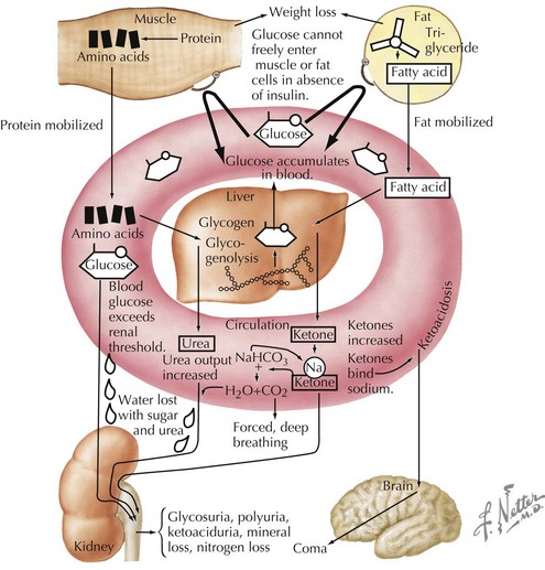
Insulin is an anabolic hormone that stimulates glucose uptake and hepatic glycogen synthesis and inhibits hepatic gluconeogenesis and glycogenolysis. It also stimulates lipogenesis, amino acid uptake, and protein synthesis (see Chapter 4, Figure 4-1). The absence of insulin triggers a series of biochemical events that emulate a starvation state even when food intake is adequate and that result in hyperglycemia and ketoacidosis (Figures 71-1 and 4-2). Glucose uptake by peripheral tissues is reduced, and hepatic glycogenolysis and gluconeogenesis are stimulated by insulin deficiency, which produces hyperglycemia. Lipolysis, proteolysis, and fatty acid oxidation lead to the accumulation of ketone bodies (β-hydroxybutyrate and acetoacetate), which eventually leads to metabolic acidosis.


As the serum glucose increases above 180 mg/dL, the renal threshold for glucose reabsorption, glycosuria results. Glycosuria causes an osmotic diuresis, resulting in polyuria and compensatory polydipsia. Over time, hyperosmolarity and dehydration develop, and decreased tissue perfusion can elicit a mild lactic acidosis. Patients without free access to fluid, such as infants and those with developmental disorders, are especially at risk. The osmotic diuresis also leads to the loss of crucial electrolytes, such as sodium, potassium, phosphorus, magnesium, and calcium. Metabolic acidosis and dehydration also stimulate counterregulatory hormones, such as growth hormone, cortisol, and epinephrine, further antagonizing insulin action. The end result is a serious metabolic disorder termed diabetic ketoacidosis (DKA; see Chapter 4).


Clinical Presentation


The clinical presentation of diabetes varies from asymptomatic hyperglycemia to life-threatening severe DKA. The majority of children with diabetes present with symptoms such as polyuria, polydipsia, nocturia, polyphagia, weight loss, dehydration, abdominal pain, vomiting, or lethargy. A history of secondary enuresis is not uncommon. Hyperglycemia and fluid compartment shifts can also affect the lens of the eye, causing blurry vision. The breakdown of protein and fat stores results in weight loss. Ketonemia can cause abdominal pain and vomiting. Pancreatitis can occur. A family history of other autoimmune diseases, such as thyroiditis and celiac disease, may be present.


Complicating the presentation is the fact that patients with new-onset diabetes often present during an intercurrent illness, which may confound the classic presentation of diabetes. In addition, a number of other conditions should be considered in the differential diagnosis of diabetes (Table 71-1).


Table 71-1 Differential Diagnosis


Symptom Differential Diagnosis
Polyuria Diabetes insipidus, urinary tract infection, psychogenic polydipsia
Polydipsia Diabetes insipidus, psychogenic polydipsia
Glycosuria Benign renal glycosuria
Weight loss Anorexia nervosa, inflammatory bowel disease, celiac disease, infectious disease
Vomiting, abdominal pain Gastroenteritis, inflammatory bowel disease, appendicitis, toxic ingestion, pancreatitis
Abnormal breathing Pneumonia, asthma exacerbation
Hyperglycemia Stress-induced hyperglycemia, medication-induced hyperglycemia

On physical examination, the respiratory status must be assessed first to determine the adequacy of the patient’s airway. Patients in DKA can present with tachypnea and deep, labored respirations called Kussmaul’s respirations. This breathing pattern occurs as respiratory compensation for the metabolic acidosis. It is important to evaluate a patient’s hydration status (tachycardia; hypotension; poor skin turgor; dry mucous membranes; sunken eyes; and in infants, sunken fontanelles), perfusion status (cool skin, delayed capillary refill), and mental status. The degree of dehydration (e.g., 5%, 10%, 20%) should be estimated, keeping in mind that intravascular fluids are preserved at the expense of intracellular fluids, and the physical examination will underestimate the degree of dehydration. Any abnormal cardiac findings should prompt immediate evaluation because electrolyte abnormalities in DKA can cause life-threatening arrhythmias. It is essential to fully evaluate neurocognitive status, with particular emphasis on mental status, because of the risk of cerebral edema.


The physical examination can disclose additional significant findings, such as a fruity breath odor secondary to ketoacidosis, candidal infections (particularly in the genital area) resulting from hyperglycemia, and nasopharyngeal infection (rhinocerebral mucormycosis). Pubertal status should be noted. Patients should also be examined for evidence of other autoimmune disorders, such as thyroiditis (goiter) and Addison’s disease (hyperpigmentation).


< div class='tao-gold-member'>

Stay updated, free articles. Join our Telegram channel

Jun 19, 2016 | Posted by in PEDIATRICS | Comments Off on Diabetes Mellitus

Full access? Get Clinical Tree

Get Clinical Tree app for offline access