122 Dermatologic Morphology
Basic Structure Of The Skin
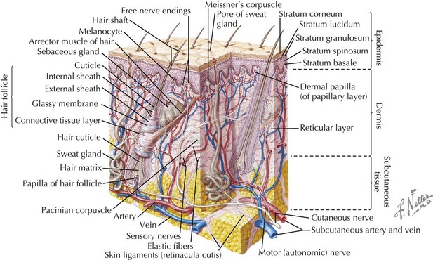
The skin has three basic layers: the epidermis, dermis, and subcutaneous tissue (Figure 122-1). Throughout these layers are additional structures and appendages that contribute to the skin’s functionality.
Epidermis
The outermost layer of the skin, the epidermis, is composed primarily of keratinocytes to form a stratified epithelial tissue that functions primarily to protect against the external environment and prevent water loss. The innermost layer of the epidermis consists of the basal cells at the dermal–epidermal junction. Basal cells divide to form keratinocytes, which undergo an evolution by initially flattening out to form the stratum granulosum. The keratinocytes eventually die and form the outermost barrier, the stratum corneum, which is continually replenished. The epidermis also has melanocytes, the melanin-producing cells of the skin, Langerhans’ cells, which are dendritic cells derived from macrophages and perform immune surveillance, as well as Merkel’s cells, which are mechanosensory touch receptors.
Dermis
The dermis is composed primarily of a matrix of fibroblasts that produce the support structure of the skin by synthesizing collagen and elastic fibers. The part of the dermis closest to the epidermis is the more cellular layer, the papillary layer. The papillary dermis supports unmyelinated nerve endings that transmit sensations of pain, itch, and temperature. Deep to this is the reticular layer, which is dense in collagen and elastic fibers. Both layers contain blood and lymphatic vessels. In addition, the dermis supports most of the skin’s appendages, including the hair follicle, apocrine and eccrine sweat glands, and sebaceous glands.
Subcutaneous Tissue
The subcutaneous tissue of the skin serves to conserve body heat and as a protective cushion. The subcutaneous tissue is composed primarily of adipocytes, which play important roles in glucose and fat metabolism. In addition, factors released by adipocytes contribute to wound healing, vascular remodeling, and the inflammatory and immune responses. A fibrous network anchors the adipocytes to deeper structures, such as muscular fascia and periosteum.
Approach To Dermatologic Morphology And Disease
Recognition of cutaneous lesions begins with basic understanding of dermatologic terminology and morphology. As with any other disease process, diagnosis of cutaneous disease begins with a thorough history and physical examination. A thorough examination includes careful inspection of the body surface, including the mucous membranes, nails, and hair. The differential diagnosis is guided by the distribution and configuration of lesions. More careful examination of individual lesions, including inspection and palpation, helps identify the primary lesion. The primary lesion is defined as the basic, most representative lesion. Lesions often undergo secondary changes as a result of scratching, infection, or treatment. Identification of the primary lesions allows accurate description and aids in generation of a differential diagnosis.
Primary Lesions
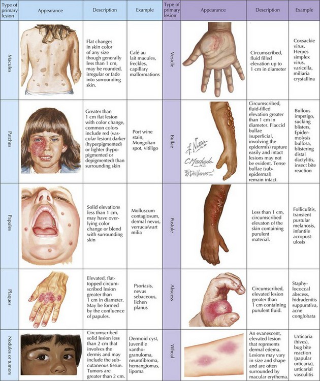
Primary lesions (Figure 122-2) are characterized by their diameter and depth. A macule is a flat lesion that can be seen by changes in skin color but cannot be felt. The border may be well circumscribed or may gradually blend into the surrounding skin. It may be of any size, but the term is generally used to describe lesions smaller than 1 cm. Flat lesions larger than 1 cm are termed patches. Similar to macules, papules are small (<1 cm) lesions but are palpable with the greatest mass above the surface of surrounding skin. Larger elevated skin lesions are termed plaques. Plaques may be formed by a confluence of papules or can be the primary lesion. Palpable, solitary lesions whose mass is primarily below the surface, in the dermis and subcutaneous tissue, are termed either nodules (0.5-2 cm) or tumors. Tumors may be benign or malignant.

Stay updated, free articles. Join our Telegram channel

Full access? Get Clinical Tree

