34 Dentition and Common Oral Lesions
Dentition
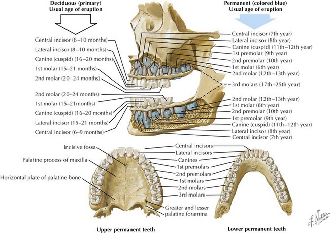
The development of the teeth begins in utero and continues well into adolescence. The 20 primary teeth (also known as deciduous or milk teeth) typically erupt between the ages of 6 months and 2 years. The exfoliation of the primary dentition and the eruption of the 32 permanent teeth usually begin at around age 6 years. On each side of the mouth, the mature permanent dentition consists of maxillary and mandibular central incisors, lateral incisors, canines, two premolars, and three molars (Figure 34-1).
Normal Tooth Anatomy
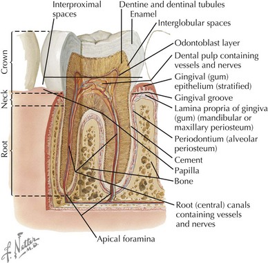
The visible portion of the tooth, which protrudes from the gingiva, is the crown, and its hard surface is an enamel made of hydroxyapatite crystals. The portion of the tooth that connects to the maxillary and mandibular alveolar bones is the root, and its hard covering is called cement. The neck of the tooth is the portion that connects the crown and the root. The periodontal ligament, or periodontal membrane, holds the root in place by attaching the cement to the periosteum of the alveolar bone. Beneath the protective overlying enamel and cement, a layer known as dentin provides the bulk of the body of the tooth. The innermost portion of the tooth, surrounded and protected by dentin, is the pulp chamber, which contains nerves, blood vessels, and connective tissue (Figure 34-2).
Dental Trauma
The most common traumatic injuries in pediatric dentistry are luxation injuries to the maxillary central incisors followed by the maxillary lateral incisors and mandibular incisors. Luxation injuries range from simple concussion, in which the tooth and ligament may be injured without being displaced or knocked loose, to avulsion, in which the tooth in its entirety is displaced from the socket. They also include intrusion, extrusion, subluxation, and lateral luxation injuries in which the tooth may be displaced in any direction with varying degrees of injury to the periodontal ligament.
The most common mechanism of injury for dental trauma is falls, especially in toddlers who are learning to walk. Fractures are also common sequelae of dental trauma, and they may be uncomplicated (involving only enamel or enamel and dentin) or complicated (involving pulp) and involve the crown or the root. Dental fractures are more common in boys and in children whose maxillary teeth more substantially override the mandibular teeth.
A thorough physical examination of the mouth in dental trauma should reveal evidence of soft tissue injury (to the lips, frenula, tongue, buccal and lingual mucosa, hard and soft palate); fracture of the teeth (with attention to whether enamel, dentin, or pulp is exposed); loose, displaced, or missing teeth; pain, tenderness, or sensitivity; or malocclusion. The clinician must always consider the possibility of child abuse and be alert for suspicious signs such as bruising or a torn upper labial frenulum. Radiographic imaging may be appropriate to reveal fractures to the teeth and supporting bone or to locate missing tooth fragments (which may have been swallowed, aspirated, or completely intruded into the alveolar socket).
The focus of management in dental trauma is to prevent aspiration, infection, and injury to the permanent dentition. In injuries of the primary teeth, children with fractured, loose, or severely displaced teeth should be referred for immediate dental management; for most, however, routine follow-up is appropriate. For children with injuries to permanent teeth, maintaining the viability of the periodontal ligament is of paramount importance; thus, most children with luxation injuries of the permanent teeth require immediate referral to a pediatric dentist. In the case of an avulsed permanent tooth, the viability of the tooth is inversely proportional to the time to reimplantation. Parents should be advised to handle the tooth by the crown, gently rinse it with tap water or saline, place it back into the socket, and ask the child to maintain pressure with a finger or by biting on gauze or a clean cloth to keep the tooth in place. Transporting the tooth in milk or Hank’s balanced salt solution will also keep the tooth viable.
Dental Infections
Caries
Dental caries remains a highly prevalent disease among children, both in the primary and permanent dentition. The risk of early childhood caries is increased in babies who sleep with bottles, who graze (rather than eating at discrete mealtimes), and whose parents have untreated caries.
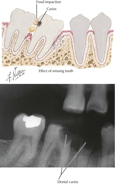
Bacteria, especially the Streptococcus mutans group, are typically transmitted from mother to infant soon after the eruption of the primary teeth. These bacteria colonize tooth surfaces and form plaques above and below the gingival margin. Caries result when these bacteria ferment sucrose from ingested dietary carbohydrate, producing organic acids on the tooth surface. These acids lead to the destruction of the hydroxyapatite crystals that give the enamel its structure, making the enamel more and more porous and eventually causing breakdown of the tooth surface (Figure 34-3). These areas of breakdown can erode through the enamel and dentin into the pulp, where an inflammatory response raises the pressure in the pulp chamber and can cause compression, and thus ischemia, of the pulp vessels. This is known as pulp necrosis.

Stay updated, free articles. Join our Telegram channel

Full access? Get Clinical Tree

