Demyelinating Diseases

78 Demyelinating Diseases


Demyelinating diseases are acquired autoimmune disorders affecting the central nervous system (CNS) in children and adolescents. Presenting with acute neurologic symptoms, these disorders are often difficult to distinguish from each other because of considerable overlap in the clinical presentation, paraclinical tests (e.g., cerebrospinal fluid [CSF] analysis), and neuroimaging features. Although some disorders, such as acute disseminated encephalomyelitis (ADEM), can be self-limited with a generally favorable prognosis, other demyelinating diseases, including multiple sclerosis (MS) and neuromyelitis optica (NMO), are chronic relapsing conditions that are potentially disabling.


Clinical Presentation and Differential Diagnosis


Acute Disseminated Encephalomyelitis


Historically, ADEM was defined by the development of neurologic symptoms after a vaccination or a viral or bacterial infection. In 2007, an International Pediatric MS Study Group (IPMSSG) proposed working definitions for demyelinating disorders in children. ADEM is currently defined as a first demyelinating or inflammatory event in which the child is polysymptomatic and encephalopathic. Encephalopathy can be mild (e.g., confusion or irritability) but must be present to distinguish ADEM from a clinically isolated syndrome (described below). Although a preceding illness or infection is identifiable in approximately 75% of children (with an average latency between the febrile illness and neurologic symptoms of 7-14 days), such an event is not required for the diagnosis. Magnetic resonance imaging (MRI) of the brain typically reveals multiple large (>1-2 cm) asymmetric lesions affecting the supra- and infratentorial white matter that are easily visualized using T2-weighted and fluid-attenuated inversion recovery (FLAIR) sequences. Symmetric gray matter involvement of the thalami and basal ganglia and confluent spinal cord lesions have also been described in ADEM. CSF analysis may demonstrate an elevated white blood cell (WBC) count or protein or the presence of oligoclonal bands, although the latter is more frequent in MS. The presence of encephalopathy, a preceding illness or vaccination, large asymmetric white matter lesions on MRI, symmetric gray matter involvement, and CSF WBC greater than 50 cells/mm are highly suggestive of ADEM rather than a first attack of MS. The clinical and radiographic involvement may fluctuate for the first 3 months. Thereafter, relapses may occur in the same (recurrent ADEM) or a different (multiphasic) CNS site but must meet the diagnostic criteria for ADEM described above to differentiate these conditions from MS.


Clinically Isolated Syndrome
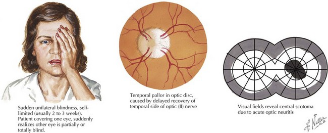

The IPMSSG defined a clinically isolated syndrome (CIS) as a first demyelinating event that may be monofocal or multifocal in the absence of encephalopathy (with the exception of a brainstem lesion, which may result in altered mental status, lethargy, or coma). Whereas the name suggests a single event, these disorders may represent the initial presentation of MS.


Optic Neuritis


Optic neuritis is characterized by acute or subacute visual loss, altered color vision, periorbital pain that is exacerbated by eye movements, and visual field defects. The neuro-ophthalmologic examination may reveal a relative afferent pupillary defect (in unilateral cases) or optic disc edema (Figure 78-1). MRI of the orbits often reveals T2/FLAIR abnormality in the optic nerve or chiasm with or without enhancement using gadolinium. Visual recovery from idiopathic optic neuritis is excellent for most children, especially in the absence of alternate diagnoses such as NMO.


Transverse Myelitis


Transverse myelitis (TM) is characterized by acute bilateral sensory, motor, or autonomic dysfunction localizable to the spinal cord. The distribution is typically asymmetric; however, this is not required for the diagnosis. Neurologic symptoms present rapidly, within hours to days, often beginning with sensory symptoms. Weakness, urinary dysfunction, and pain frequently occur depending on the extent and location of disease. In 2002, the Transverse Myelitis Consortium Working Group proposed diagnostic criteria for TM. According to these guidelines, inflammation in the spinal cord must be demonstrated by CSF analysis (pleocytosis or elevated immunoglobulin G [IgG] index) or MRI (gadolinium enhancement), which helps differentiate noninflammatory causes of spinal cord disease (including previous radiation, anterior spinal artery thrombosis, tumor, syrinx, and compressive myelopathy) from idiopathic TM. However, these criteria have not been validated in children, and normal CSF profiles have been reported. If inflammation is not detected in the CSF or on MRI, the Working Group recommends a repeat spinal tap or MRI 2 to 7 days after symptom onset to further look for signs of inflammation. In addition, idiopathic TM should be distinguished from symptomatic TM caused by CNS infections (e.g., Lyme disease, HIV, human T-lymphotropic virus-1, syphilis, Mycoplasma, herpes simplex virus types 1 and 2 [HSV-1 and HSV-2], HHV-6, varicella zoster virus, EBV, cytomegalovirus, and enteroviruses) or systemic diseases (e.g., connective tissue disorders such as systemic lupus erythematosus, Sjögren’s disease, sarcoidosis, or antiphospholipid antibody). Other than the presence of gadolinium enhancement, there are no proposed diagnostic criteria for the size or extent of the lesion upon neuroimaging of the spine. Nevertheless, whereas TM is often associated with longitudinally extensive lesions (greater than three or more vertebral segments), discrete lesions are more common in the initial presentation of MS. At the time of the development of TM, the presence of T2 or FLAIR abnormalities on an MRI of the brain is suggestive of a first attack of MS. The prognosis for full recovery is less favorable than for the other demyelinating diseases because some children may have residual disability in ambulation, sensation, or bladder function.

< div class='tao-gold-member'>

Stay updated, free articles. Join our Telegram channel

Jun 19, 2016 | Posted by in PEDIATRICS | Comments Off on Demyelinating Diseases

Full access? Get Clinical Tree

Get Clinical Tree app for offline access