Fig. 11.1
Rigid cystoscope
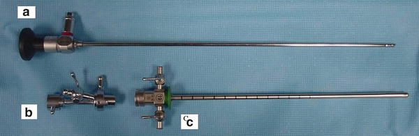
Fig. 11.2
Components of rigid cystoscope (a) telescope (b) bridge (c) sheath
Flexible cystoscopes have the ability to combine the optical system along with irrigation capabilities into a single unit. The flexible tip of the cystoscope allows for a deflection of 290° and has a diameter ranging from 15 to 18 French. See Figs. 11.3, 11.4, and 11.5. Multiple investigations have been performed comparing rigid versus flexible cystoscopy, however some studies included male patients and other studies evaluated quality-of-life outcomes between patients who underwent office flexible cystoscopy compared to those who underwent a rigid cystoscopy under general anesthesia [6]. More recently, studies by Gee et al. and Quinoz et al. comparing rigid and flexible in-office cystoscopy specifically in women, found no difference between either modality in regards to tolerance of the procedure and both techniques having equivalent morbidity [7, 8]. Also, flexible and rigid cystoscopes have been shown to be equally efficacious in their diagnostic capabilities [9]. Therefore, one should select the cystoscope that they are most comfortable with.
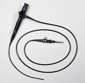
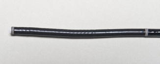

Fig. 11.3
Flexible cystoscope
Fig. 11.4
Tip of flexible cystoscope
Fig. 11.5
Tip of flexible cystoscope with maximum curvature
Illumination can be provided by either a halogen or xenon lamp along with fiberoptic or fluid-filled light cables that attach to the telescope at the eyepiece. A standard xenon 175-W lamp is the most commonly used given its immediate responsiveness once turned on and the ability to produce a high intensity illumination. Fluid-filled cables tend to have a longer shelf life, but fiberoptic cables are typically less expensive and are more susceptible to damage with routine use. Routine cystoscopy may also be performed looking through the telescope alone, but with the use of a camera and video monitoring system, patients and physicians are able to view the internal anatomy at the same time. This allows for enhanced teaching, image documentation, and potential patient distraction during the procedure [3]. Of note, complete systems are able to be purchased that routinely include a light source, camera, and video monitor.
Distending media may be either a conductive fluid (lactated ringers, normal saline), a nonconductive fluid (sterile water, 5 % glycine, 5 % mannitol, or 3 % sorbitol), or gas. Carbon dioxide and air were among the first agents used to distend the bladder during the early development of cystoscopy [4]. As technology progressed, with the potential concern of development of an “air embolism” accompanied by patient’s tolerability of the procedure and the poor visualization noted with gas cystoscopy, there was a decline in the use of air as a distending media and liquid media became the current accepted practice [4]. Selection of either a conductive or nonconductive fluid is based on whether a potential for an energy source to be used during the procedure. For diagnostic purposes, most physicians elect for either isotonic normal saline or sterile water.
Procedure in Detail
Office cystoscopy for female patients is usually performed in the dorsal lithotomy position with the patient undressed from the waist down and covered in a sterile drape. Prior to this, the patient is instructed to void and a dip-stick urinalysis is performed to rule out an active urinary tract infection. Per the American Urological Association guidelines, routine antibiotic prophylaxis is not indicated for diagnostic cystoscopy unless the following risk factors are present and a negative urine culture is not documented prior to the procedure: immunocompromised state, known anatomic abnormalities of the urinary tract, active infection, chronic steroid use, or external catheter use [1]. Consent for cystoscopy is obtained and the urethra is cleansed with either a povidine–iodine solution or equivalent antimicrobial solution if an allergy exists. A disposable urinary catheter is inserted to obtain a post void residual if indicated and a topical analgesic may or may not be applied directly into the urethra from a tube or needleless syringe.
Under sterile technique, the equipment is assembled with the proper cystoscope, a light source, distending media, and imaging source if desired. If rigid cystoscopy is selected, a 30° or 70° cystoscope is routinely used along with a 17 French sheath. For rigid cystoscopy, ensure closure of all the appropriate ports to prevent leakage, turn on the light source and open the distending media flow to allow passage of a minimal amount of liquid media. For both flexible and rigid cystoscopy, gently guide the tip of the cystoscope into the distal urethra under direct visualization and follow the central opening past the urethrovesicular junction (UVJ) into the bladder cavity. Some physicians prefer the use of a blunt obturator for the cystoscopy sheath rather than direct visualization. One may then increase the flow of the distending media to adequately distend the bladder, typically around 300 cc, or the maximum capacity tolerated by the patient.
Stay updated, free articles. Join our Telegram channel

Full access? Get Clinical Tree


