Congenital Anomalies and Benign Conditions of the Uterine Corpus and Cervix

Chapter 19 Congenital Anomalies and Benign Conditions of the Uterine Corpus and Cervix


Benign conditions of the uterine corpus and cervix are commonly encountered in gynecologic practice because they affect a woman’s fertility and can cause abnormal uterine bleeding or pelvic pain. In this chapter, congenital anomalies, benign neoplasms, epithelial changes, and functional disorders of the uterus (corpus and cervix) are discussed along with conventional and emerging therapies.


image Congenital Anomalies of the Uterine Corpus and Cervix


The upper vagina, cervix, uterine corpus, and fallopian tubes are formed from the paramesonephric (müllerian) ducts. The absence of a Y chromosome and the resultant absence of müllerian inhibiting substance lead to the development of the paramesonephric system, with the regression of the mesonephric system. The paramesonephric ducts first arise at 6 weeks’ gestation lateral to the cranial pole of the mesonephric duct and expand caudally. By 9 to 10 weeks, they fuse in the midline at the urogenital septum to form the uterovaginal primordium. Later, dissolution of the septum between the fused paramesonephric ducts leads to the development of a single uterus and cervix.


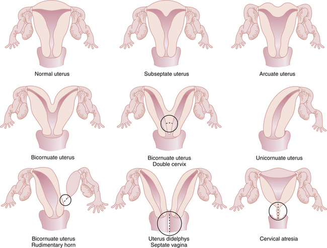
The most common anomalies of the uterus result from either incomplete fusion of the paramesonephric ducts, incomplete dissolution of the midline fusion of those ducts, or formation failures. Figure 19-1 shows variations of uterine and cervical development and demonstrates that communication between the dual systems can exist at several levels. Failure of fusion is most evident in uterus didelphys, which presents with two separate uterine bodies, each with its own cervix and attached fallopian tube and vagina. A bicornuate uterus with a rudimentary horn also represents a fusion failure. Less complete fusion failure is seen in the bicornuate uterus with or without double cervices. Incomplete dissolution of the midline fusion of the paramesonephrica explains the septate uterus. Failure of formation can be seen in the unicornuate uterus. In müllerian agenesis, there is complete lack of development of the paramesonephric system. The affected woman generally has incomplete development of the fallopian tubes associated with the absence of the uterus and most of the vagina. All these conditions occur in normal karyotypic and phenotypic females but can be associated with important anomalies of the urinary system such as a horseshoe or pelvic kidney.


The most common congenital cervical anomalies are the result of malfusion of the paramesonephric (müllerian) ducts with varying degrees of separation, as seen in the didelphys cervix or septate cervix.


These different anatomies may have a significant effect on a woman’s risk for infertility and early pregnancy loss and may also cause dysmenorrhea and dyspareunia. Women with fusion anomalies may present with menstrual blood trapped in a noncommunicating uterine horn or vagina.


In addition to these macroscopic differences, subtle anomalies may exist within the uterine vascular system, such as an arteriovenous malformation, rupture of which may cause life-threatening hemorrhage.


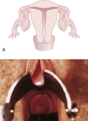
Although all these anomalies can occur spontaneously, they may also be caused by early maternal exposure to certain drugs. The most notable of these drugs is diethylstilbestrol (DES). A DES-exposed female infant has an increased risk for a small, T-shaped endometrial cavity (Figure 19-2A) or cervical collar deformity (Figure 19-2B). DES exposure in utero can also produce fallopian tube abnormalities, although it does not appear to cause abnormalities of the urinary tract.


image Benign Neoplastic Conditions


UTERINE LEIOMYOMAS


Uterine leiomyomas (“fibroids”) are benign tumors derived from the smooth muscle cells of the myometrium. They are the most common neoplasm of the uterus. Estimates are that more than 45% of women have leiomyomas by the fifth decade of life, but most are asymptomatic. However, leiomyomas can cause excessive uterine bleeding, pelvic pressure and pain, and infertility. They are the primary indication for about 200,000 hysterectomies in the United States each year. Although leiomyomas have the potential to grow to impressive sizes, their malignant potential is minimal. Sarcomatous changes occur in less than 1 per 1000 uteri with fibroids.


Risk factors for developing leiomyomas include increasing age during the reproductive years, ethnicity (African American women have at least a twofold to threefold increased risk compared with white women), nulliparity, and family history. The data are suggestive that higher body mass index is associated with a greater risk for leiomyomas. Oral contraceptive pills and depot medroxyprogesterone acetate (DMPA) injections may be associated with reduced risk.


Characteristics of Leiomyomas


Leiomyomas are usually spherical, well-circumscribed, white, firm lesions with a whorled appearance on cut section. Although the leiomyoma appears discrete, it does not have a true cellular capsule. Compressed smooth muscle cells on the tumor’s periphery provide the false impression of such a capsule. Few blood vessels and lymphatics traverse the pseudocapsule, leading to degenerative changes as the tumors enlarge. The most commonly observed degenerative change is that of hyaline acellularity, in which the fibrous and muscle tissues are replaced with hyaline tissue. If the hyaline substance breaks down from a further reduction in blood supply, cystic degeneration may occur. Calcification may occur in degenerated fibroids, particularly after the menopause. Fatty degeneration may also occur but is rare. During pregnancy, 5% to 10% of women with fibroids undergo a painful red or carneous degeneration caused by hemorrhage into the tumor.


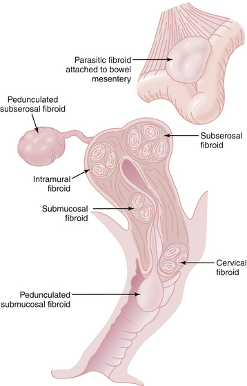
Leiomyomas always arise within the myometrium (intramural), but some migrate toward the serosal surface (subserosal) or toward the endometrium (submucosal), as depicted in Figure 19-3. Individual tumors may migrate further when they develop large pedicles. The submucosal leiomyomas can extend through the endometrial canal and abort from the cervical os. An aborting leiomyoma causes significant bleeding and cramping pain. A subserosal leiomyoma on a long pedicle can present as a mass that feels separate from the uterus. Rarely, pedunculated subserosal myomas attach to the blood supply of the omentum or bowel mesentery and lose their uterine connections to become parasitic leiomyomas. Leiomyomas can also arise in the cervix, between the leaves of the broad ligament (intraligamentous), and in the various supporting ligaments (round or uterosacral) of the uterus. Leiomyomas can invade and fill the vena cava retrograde up to the level of the atrium (intravenous leiomyomatosis).

Symptoms of Leiomyomas


Most uterine leiomyomas cause no symptoms. On occasion, the patient may complain of a lower abdominal mass if the fibroid protrudes above the pelvis. Symptomatic women may complain of pelvic pressure, congestion, bloating, a feeling of heaviness in the lower abdomen, or lower back pain. If the fibroid presses on the bladder, there may be a problem with frequency of urination. Urinary retention and hydronephrosis are rare but result from the fact that the bladder and large leiomyomas compete for space within the pelvis.


Prolonged or heavy menstrual bleeding may be associated with intramural or submucosal myoma. Intermenstrual bleeding is not characteristic of these tumors but may occasionally occur with submucous myomas ulcerating through the endometrial lining. Excessive bleeding may result in anemia, weakness, dyspnea, and even congestive heart failure.


Fibroids are not generally painful, but severe pain may be associated with red degeneration (acute infarction) within a fibroid, which occurs most commonly during pregnancy. In addition, pressure pains may occur in the lower abdomen and pelvis if a myomatous uterus becomes incarcerated within the pelvis. Dyspareunia is also common with incarceration. There is an increased incidence of secondary dysmenorrhea in women with uterine myomas, generally caused by more frequent episodes of uterine bleeding. Although many women with uterine myomas become pregnant and carry their pregnancies to term, submucosal leiomyomas may be associated with an increased incidence of infertility because of placentation challenges.


Jun 4, 2016 | Posted by in GYNECOLOGY | Comments Off on Congenital Anomalies and Benign Conditions of the Uterine Corpus and Cervix

Full access? Get Clinical Tree

Get Clinical Tree app for offline access