Congenital Anomalies and Benign Conditions of the Ovaries and Fallopian Tubes

Chapter 20 Congenital Anomalies and Benign Conditions of the Ovaries and Fallopian Tubes


The ovaries and fallopian tubes, along with the blood vessels, ligaments, and connective tissues located along both sides of the uterus, are referred to as the adnexa. In this chapter, the normal development of the ovaries and fallopian tubes is discussed to facilitate a better understanding of the workup and management of benign adnexal masses.


image Congenital Anomalies of the Ovaries and Fallopian Tubes


Abnormal embryologic development of the ovaries is uncommon. Congenital duplication or absence of ovarian tissue may occur, as may ectopic ovarian tissue and supernumerary ovaries. Although rare, the sexual bipotentiality noted in embryologic development can progress without the usual regression of one system, producing an ovotestis and subsequent intersex problems.


Genetic chromosomal disorders, such as Turner syndrome (45 XO), are associated with a lack of normal gonadal development, as evidenced by the rudimentary streaked ovaries that are a hallmark of the disorder. Women with Turner syndrome usually progress through puberty and develop secondary sexual characteristics but enter menopause shortly thereafter. This provides evidence that two X chromosomes are required for normal ovarian development. Testicular predominance occurs with the addition of a single Y chromosome, even in the face of multiple X chromosomes. Such predominance is seen in Klinefelter syndrome (47 XXY), in which testicular development occurs embryologically. In complete androgen insensitivity syndrome (46 XY), which is also known as testicular feminization, the lack of androgen receptors produces a phenotypic female in the face of a Y chromosome. The gonads in these women (functioning testes) should be removed (usually after puberty) because of their significant malignant potential.


Isolated anomalies of the fallopian tubes, the end result of abnormal development of the proximal unfused portions of the paramesonephric ducts, are rare. Aplasia or atresia, usually of the distal ampullary segment of the fallopian tube, is most commonly unilateral in the presence of otherwise normal development. Bilateral aplasia is noted in some cases of uterine and vaginal agenesis. Complete duplication of the fallopian tubes is rarely seen, but distal duplication and accessory ostia are relatively common.


In addition, women exposed in utero to certain drugs, such as diethylstilbestrol (DES), may have abnormalities in the architecture of the fallopian tubes; with DES exposure, the tubes may be shortened, distorted, or clubbed.


image Benign Conditions of the Ovaries


FUNCTIONAL AND BENIGN OVARIAN TUMORS


The human ovary has a striking propensity to develop a wide variety of tumors, most of which are benign. As indicated in Table 20-1, ovarian masses may be functional, inflammatory, metaplastic, or neoplastic. During the childbearing years, 70% of noninflammatory benign ovarian tumors are functional. The remainder are either neoplasms (20%) or endometriomas (10%). The management of ovarian tumors, whether functional, benign, or malignant, involves difficult decisions that may affect a woman’s hormonal status or her future fertility. Although only functional cysts and benign ovarian neoplasms are considered in detail in this chapter, diagnostic methods to differentiate benign masses from malignant ones are discussed.


TABLE 20-1 DIFFERENTIAL DIAGNOSIS OF OVARIAN MASSES


Pathogenesis Specific Type
Functional Follicular cysts
  Lutein cysts
  Polycystic ovaries
Inflammatory Salpingo-oophoritis
  Pyogenic oophoritis—puerperal, abortal, or related to an intrauterine device
  Granulomatous oophoritis
Metaplastic Endometriomas
Neoplastic Premenarchal years—10% are malignant
  Menstruating years—15% are malignant
  Postmenopausal years—50% are malignant


Functional Ovarian Cysts and Tumors


Dozens of ovarian follicles form the “cohort of follicles” of each menstrual cycle. To be classified a functional cyst, the follicle must reach a diameter of at least 3 cm. Functional cysts may cause pelvic pain, a dull sensation, or heaviness in the pelvis.


A follicular cyst, lined by one or more layers of granulosa cells, develops when an ovarian follicle fails to rupture. Similarly, a lutein cyst may develop if the corpus luteum becomes cystic, grows to larger than 3 cm, and fails to regress normally after 14 days. Hemorrhagic cysts, especially hemorrhagic corpus luteum cysts, are more likely to cause symptoms and are more vulnerable to rupture toward the end of the menstrual cycle. The hemorrhage within the cyst results from invasion of the ovarian vessels into the corpus luteum cyst 2 to 3 days after ovulation.


Other specific types of lutein cysts may occur with abnormally high serum levels of human chorionic gonadotropin (hCG) or increased ovarian sensitivity to gonadotropins. Theca-lutein cysts may develop in association with the high levels of hCG present in patients with a hydatidiform mole or choriocarcinoma. Patients undergoing ovulation induction with gonadotropins or clomiphene may also develop theca-lutein cysts. Theca-lutein cysts are usually bilateral, may become quite large (>30 cm), and characteristically regress slowly after the gonadotropin level falls. Rarely, when follicles are stimulated with gonadotropins, theca-lutein cysts can become so extensive as to cause massive ascites and dangerous problems with systemic fluid imbalance.


A luteoma of pregnancy is a related condition in which there is a hyperplasic reaction of ovarian theca cells, presumably from prolonged hCG stimulation during pregnancy. The luteomas characteristically appear as brown to reddish-brown nodules that may be cystic or solid. A luteoma of pregnancy (Figure 20-1) may be associated with multifetal pregnancies or hydramnios. They can cause maternal virilization in 30% of women and, less often, ambiguous genitalia in a female fetus. Although ovarian enlargement may be impressive, surgical resection is not indicated because luteomas regress spontaneously postpartum.
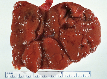


Polycystic ovary syndrome, a functional disorder generally associated with chronic anovulation and hyperandrogenism, can also produce enlarged ovaries with multiple simple follicles (Figure 20-2). The hormonal aspects of this syndrome are discussed further in Chapter 32.
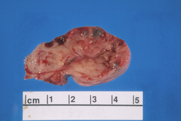


DIAGNOSIS


The presumptive diagnosis of a functional ovarian tumor is usually made when a 5- to 8-cm cystic adnexal mass is noted on bimanual examination; it is confirmed when the lesion regresses over the course of the next several cycles. In general, a functional cyst is mobile, unilateral, and not associated with ascites. On rare occasions, the mass may exceed 8 cm and be quite tender to palpation. On occasion, hemorrhagic lutein cysts may have a solid rather than a cystic consistency. A pelvic ultrasound will confirm the cystic nature of the mass, but it cannot differentiate with certainty between a functional and a neoplastic tumor.


All suspicious adnexal masses in premenopausal and postmenopausal women must be carefully evaluated. Table 20-2 contains a useful risk assessment tool, the risk for malignancy index (RMI), which can be used to help identify those ovarian masses that could be malignant. The RMI has high sensitivity (87%) and specificity (97%).


TABLE 20-2 CALCULATION OF THE RISK OF MALIGNANCY INDEX FOR AN OVARIAN MASS


Criteria Scoring System
A. MENOPAUSAL STATUS  
Premenopausal 1
Postmenopausal 3
B. ULTRASONIC FEATURES  


C. SERUM CA-125 TITER Absolute value

Risk of Malignancy Index = A × B × C. A cut-off value of 200 discriminates a benign from a malignant mass with a sensitivity of 87% and a specificity of 97%.

Stay updated, free articles. Join our Telegram channel

Jun 4, 2016 | Posted by in GYNECOLOGY | Comments Off on Congenital Anomalies and Benign Conditions of the Ovaries and Fallopian Tubes

Full access? Get Clinical Tree

Get Clinical Tree app for offline access