11.4 Congenital and perinatal infections
Introduction
Infections in the fetus and newborn (perinatal infections) may be acquired in utero (congenital infection), around the time of delivery or in the neonatal period.
Modes of acquisition of infection
The route by which the fetus or newborn acquires the infection has important implications for management during pregnancy and the neonatal period, and for the development of appropriate intervention strategies to prevent mother-to-child transmission.
The outcome of perinatal infection may include particular constellations of congenital abnormalities, spontaneous abortion or stillbirth, or acute neonatal infection. Common clinical manifestations include:
For some congenital infections, there may be no symptoms or signs in the neonatal period and it may be weeks, months or even years before the effects first become evident.
Many organisms can cause infection in the fetus and newborn. Box 11.4.1 lists some of the more common or clinically significant.
Risk assessment
The risk of fetal damage can be estimated, based on:
• the likelihood of maternal exposure and infection
• the likelihood of transmission to the fetus
• the stage in gestation at which infection occurs – this influences the risk of vertical transmission and/or the fetal or perinatal consequences.
Only a small number of exposed infants are infected and, of these, a minority will have adverse effects. Many infections that can damage the fetus are mild or asymptomatic in the mother, and diagnosis may depend on routine antenatal screening. Whether this is appropriate depends on the frequency and severity of fetal or neonatal disease and the availability of a suitable screening test and effective intervention.
Congenital infections
• Some common viral infections (e.g. cytomegalovirus, parvovirus, rubella, varicella-zoster virus) can cause fetal infection with severe consequences if acquired during pregnancy by a non-immune woman.
• Ensuring that a woman has antenatal testing and is immunized with all recommended vaccines is an important part of care during pregnancy.
• Management of a pregnant woman exposed to relevant viral infections depends on a careful risk assessment, which includes knowledge of the woman’s immune status, history of the exposure, stage in the pregnancy, and clinical presentation in the woman.
Organisms associated with perinatal infection
Cytomegalovirus
Primary cytomegalovirus (CMV) infection is usually asymptomatic or causes a non-specific illness with fever, atypical lymphocytosis and mild hepatitis. The virus remains in a latent state with periodic asymptomatic reactivation and excretion in urine, saliva or genital secretions. Primary maternal infection or reactivation can result in fetal CMV infection, although fetal damage is more likely to be associated with primary infection. CMV can infect the fetus transplacentally to cause congenital infection.
Cytomegalovirus can also be transmitted during or after delivery when the neonate comes in contact with maternal genital secretions or with breast milk. However, it appears that there are no hearing or neurodevelopmental sequelae.
Congenital CMV affects approximately 3–12 per 1000 births and causes 10–30% of childhood sensorineural hearing loss (SNHL). Infants with asymptomatic congenital CMV have a risk of developing hearing loss or intellectual disability. Early intervention can minimize the impact of SNHL on language development. Moreover, antiviral treatment may improve outcome.
The best evidence for primary maternal infection is seroconversion but this may not be demonstrable if investigation is delayed. Specific immunoglobulin (Ig) M may indicate recent infection but is unreliable: it may be detectable for months, can rise after reactivation, and false-positive results are not uncommon.
Primary infection
• 50% of young women are seronegative (susceptible). In developing countries and lower socioeconomic groups, primary infection occurs at a younger age and fewer women are susceptible.
• 1% of women seroconvert during pregnancy.
• 30% of fetuses of women with primary infection are infected.
• The risk of congenital CMV infection after primary maternal CMV infection remains increased for up to 4 years after seroconversion, with the highest risk being in the first 2 years.
• Infection is transplacental; severe fetal damage is more likely early in gestation.
• 10% of infants infected during primary maternal infection are symptomatic at birth: of these, 90% have significant long-term handicap.
• 90% of infants infected during primary maternal infection are asymptomatic at birth: of these, 10% go on to develop deafness or intellectual handicap.
• The overall incidence of congenital infection due to primary maternal infection is 1 in 1000.
Reactivation
• 20–30% of seropositive women reactivate latent infection during pregnancy.
• 2–5% of their infants are infected in utero but significant CMV disease is rare; mild sequelae (unilateral deafness) occur infrequently (< 10%).
• The overall incidence of congenital infection is 1–2% – the majority are unaffected.
Clinical features
The clinical features of severe intrauterine CMV infection include:
• intrauterine growth restriction
• hepatosplenomegaly, hepatitis
• microcephaly, encephalitis, cerebral calcification and chorioretinitis
• SNHL in congenital CMV affects 50% of symptomatic infants and 10% of asymptomatic infants. It is the most common long-term consequence. It ranges from mild unilateral to profound bilateral hearing loss. In asymptomatic infants it may be underdiagnosed because the SNHL is detected too late to prove congenital infection
• cerebral palsy, intellectual disability, epilepsy and visual impairment.
Diagnosis
If primary maternal infection is suspected or cannot be excluded, fetal infection can be diagnosed by culture and/or polymerase chain reaction (PCR) analysis of amniotic fluid at 18 weeks’ gestation. Depending on when maternal infection occurred, the risk of fetal damage may justify termination of pregnancy.
Congenital CMV infection is diagnosed by a positive culture of urine or saliva collected in the first 3 weeks of life; after that it is difficult to distinguish congenital from postnatal infection. Testing for CMV IgM in the infant’s serum is less sensitive and specific. Congenital infection may be confirmed by PCR analysis of dried blood spots (e.g. Guthrie card).
Treatment
Antiviral drugs active against CMV are contraindicated during pregnancy but have been used with limited success in congenitally infected infants. Intravenous ganciclovir and oral valganciclovir have been shown to reduce hearing deterioration in infants with symptomatic congenital CMV and central nervous system involvement. However, use of these medications is limited by their side-effects. There is currently no indication for systemic antiviral therapy for infants with asymptomatic infection at birth.
A 32-year-old woman delivered a small-for-gestational-age baby boy weighing 2400 g at 38 weeks’ gestation. His head circumference was smaller than expected for his weight and he had hepatosplenomegaly. He also had thrombocytopenia.
Rubella, CMV, Toxoplasma and syphilis serology were performed on the baby and compared with the mother’s antenatal serological results. The baby had urine collected for viral culture. The baby had CMV IgG and IgM present, and CMV was cultured from the urine, confirming congenital CMV infection.
The potential side-effects of 6 weeks of intravenous ganciclovir therapy in this child must be weighed against a possible small benefit in terms of reduction of hearing deterioration. Affected neonates should have baseline audiology testing, and this should be repeated in the first 6 months of life. They should also be monitored for developmental delay.
Parvovirus B19
Most cases of parvovirus B19 infection are asymptomatic. The most common clinical presentation of infection is erythema infectiosum, or ‘slapped cheek disease’ in children.
Parvovirus B19 can also cause:
• mild respiratory tract illness without rash
• chronic bone marrow failure in immunodeficient patients
• transient aplastic crisis in patients with haemolytic anaemia (e.g. sickle cell disease).
Approximately 60% of adult women are immune. The risk of infection in seronegative women is greatest in women exposed to an infected child at home (approximately 50%). The risk for childcare and primary school teachers exposed is 20–30%, and the risk overall depends on exposure to children but is approximately 10–20%.
Fetal risks following maternal infection
In 50% of cases of maternal infection, the fetus is unaffected. Fetal damage occurs only if maternal infection occurs before 20 weeks’ gestation. The complications include:
• fetal loss in the first 20 weeks of pregnancy (15% compared with 5% in controls, i.e. 10% excess fetal loss)
• congenital abnormalities: < 1% (anecdotal reports only)
• hydrops fetalis: following maternal infections at 9–20 weeks’ gestation (incidence is approximately 3%); may result in:
Table 11.4.1 Risk assessment – parvovirus B19
Any pregnant woman exposed to parvovirus (%) | Pregnant woman with proven recent infection (%) | |
---|---|---|
Excess fetal loss in first 20 weeks | 0.4–1 (1 in 100–250) | 5 (1 in 20) |
Death from hydrops or its treatment | 0.05–0.1 (1 in 850–2000) | 0.6 (1 in 170) |
There are no specific congenital abnormalities associated with maternal parvovirus infection.
Diagnosis
Diagnosis in children is mainly clinical. In potentially infected women, serology and PCR can be performed. IgM is detectable within 1–3 weeks of exposure and usually remains detectable for 2–3 months. Specific IgG may rise 2–3 weeks following infection. Parvovirus DNA can be detected in serum for up to 9 months after the acute viraemic phase in some patients, so it does not necessarily indicate acute infection.
Management
Pregnant school teachers or childcare workers do not need to be excluded from work, even during an epidemic (nor do infected children). It is certainly not practicable to prevent exposure at home. Pregnant women who have been exposed to parvovirus, and those with an illness consistent with parvovirus, should be tested serologically. If maternal infection is confirmed, the pregnancy should be monitored with serial ultrasonography.
A 24-year-old primary school teacher is 12 weeks’ pregnant. She has a 3-year-old who is well at the moment. She is concerned because there is a boy in her class with ‘slapped cheek disease’. What advice should be given to her?
The likelihood is that she is immune – 60% of women of child-bearing age are seropositive. She should have her serology done to check. Even if she is not immune, there is no point in her staying away from school. The boy with slapped cheek disease is probably no longer infectious – once the rash is apparent children are not infectious, and there may well have been others in the class with asymptomatic infection. If there have been many cases of parvovirus infection around, her 3-year-old may have had it too, and the greatest risk of infection for a pregnant mother is from her own children.
If the woman is seronegative when checked, she should be offered repeat serology in 2 weeks. If the serology is negative at this time, nothing further needs to be done. If she has evidence of seroconversion, there is a 50% chance that the fetus will be infected and a very small risk that she will lose the pregnancy or that the fetus will develop hydrops. She should have ultrasonography at 1–2-week intervals for the next 8 weeks, checking for the development of fetal hydrops. Even if this develops, at least 60% of fetuses will have a good outcome.
Rubella
The teratogenic effects of rubella were first noted in 1941 by an Australian ophthalmologist, who recognized several cases of congenital cataract following a large outbreak of rubella. Maternal rubella is now rare in many industrialized countries with rubella vaccination programmes. However, in many developing countries, congenital rubella syndrome remains a major cause of developmental anomalies, particularly blindness and deafness.
Clinical features
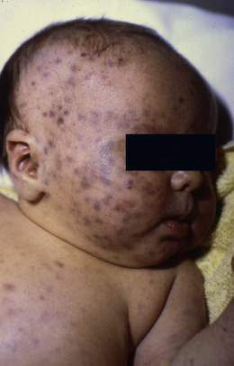
The risk of fetal infection and damage is greatest during the first 8 weeks of pregnancy and damage is rare after 16 weeks. Congenital rubella syndrome (Fig. 11.4.1) may include a number of clinical features, some of which may not present until adolescence or adulthood:
• intrauterine growth restriction
• neonatal purpuric rash and hepatosplenomegaly
• microcephaly and developmental delay
• cardiac: pulmonary artery hypoplasia, patent ductus arteriosus
• eye: cataract, retinopathy; microphthalmia
Diagnosis
Congenital rubella is confirmed by:
• isolation of rubella virus from saliva, tears, urine, cerebrospinal fluid (CSF) or tissue during the first 3 months of life
• demonstration of specific IgM antibody or persistence of IgG antibody beyond 6 months of age.

Stay updated, free articles. Join our Telegram channel

Full access? Get Clinical Tree