chapter 16 Clinical Endocrine Evaluation
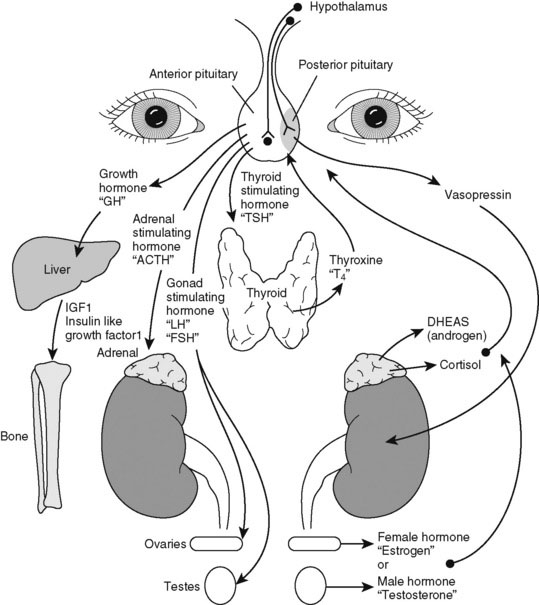
Pediatric endocrinology has special appeal because most childhood endocrine disorders are treatable. A specific diagnosis is usually possible on clinical grounds alone. Do not be intimidated by the myriad of hormone assays and endocrine tests needed to evaluate excesses or deficiencies. We have neatly arranged them all in an interrelated hormone map, which shows the feedback control of hormone action (Fig. 16–1). A good practice is to draw such a picture as you are explaining the details of hormone action to the family. Doing so slows down your explanation and makes you translate the medical lingo while still providing the correct names for hormones.

FIGURE 16–1 A map of the endocrine system. Draw the appropriate portion of the map as you sit with the family and the child. It is a way to explain hormone deficiencies or excesses. Tell the family that they are at the same stage as a first-year medical student, getting their first lecture in endocrinology. Tailor the drawing to the specific disorder, however, to avoid giving them the whole lecture.
Probably the most common endocrine complaints that parents express concern a child who is too small, too heavy, too thin, or too tall or whose sexual development is premature or delayed. On first hearing such complaints, listen; do not be judgmental. Even if there is no serious disease, the family or child needs a careful assessment and a clear explanation. Be prepared to spend time helping individuals understand and accept the many variations of normal development, and be sure to talk directly to the child about his or her self-perception and future prospects for growth and development.
Chief Characteristics of the Child with Growth Hormone Deficiency
Most short children do not have growth hormone deficiency (GHD), but because GHD is so highly treatable, it must be carefully considered and excluded. In children, GHD may be organic (tumor, cranial irradiation, or congenital malformation) or idiopathic. Hypothalamic-pituitary failure of growth hormone (GH) secretion can occur alone or in combination with deficiencies of one or several pituitary hormones. Certain midline defects are often associated with GHD, including cleft palate, choanal atresia, a single upper central incisor, and optic nerve hypoplasia (with or without an absence of the septum pellucidum, which is revealed by magnetic resonance imaging [MRI]). A classic MRI finding associated with GHD consists of an ectopic posterior pituitary bright spot, an absent or attenuated stalk, and a small anterior pituitary. Recent advances in molecular biology have shown that many cases of GHD with or without midline defects that were previously thought to be sporadic, actually have a specific genetic basis.
In examining children who are unusually short, always ask about a history of symptoms that may suggest hypoglycemia in infancy and early childhood. Because GH is a major counterregulatory hormone to insulin, deficiency of this hormone makes some children susceptible to hypoglycemia, particularly during times of fasting or during mild illness when food intake is marginal.
When asked about symptoms of hypoglycemia, parents of a child with GHD may describe episodes in which the child was found limp, glassy-eyed, stuporous, and possibly unresponsive to verbal commands. Most parents have successfully tried the natural cure, fruit juice or its equivalent. If the child is left untreated, convulsions may result, bringing the child to immediate medical attention. Unlike febrile seizures or epilepsy, a hypoglycemic episode rarely ends in spontaneous recovery without administration of sugar.
Parents of a child with idiopathic or early childhood GHD usually report that the child has always been smaller than others but that the difference became more noticeable when he or she started school. Often, no height record is available, so always ask how frequently the child’s shoe and clothing sizes have changed.
New-onset growth failure with GHD in an older child is more likely due to a destructive lesion, such as a tumor or previous cranial irradiation. Assessment of a child with short stature at any age must include careful questions about headaches.
Headaches as a result of a tumor often occur without a precipitating cause, at night or in the early morning, and may or may not be accompanied by vomiting. It is rare for children or adolescents in whom physical examination detects optic atrophy or a visual field defect to have recognized a visual problem themselves. Ask whether they have felt an increase in thirst and urination, which may suggest diabetes insipidus.
Case History 1
History. Terry, age 16 months, whose parents say he has had an uncontrollable thirst and a constantly wet diaper from early infancy, is placed on the floor in the examining room. There are four bottles of water in his toy bag. Before removing his toys, he takes all the bottles out of the bag and positions them in the corners of the room. What is the likely cause? What other questions should be asked?
Diabetes insipidus is generally classified as central (as a result of destruction or maldevelopment of the hypothalamic-pituitary vasopressin axis) or nephrogenic (e.g., congenital X-linked variety usually the result of a mutation in the vasopressin V2 receptor in the kidney, as illustrated in the case history). Terry did have a family history of excessive water drinking in his male relatives, making the diagnosis nephrogenic (not pituitary) diabetes insipidus.
Approach to the Physical Examination of a Child with Possible Growth Hormone Deficiency
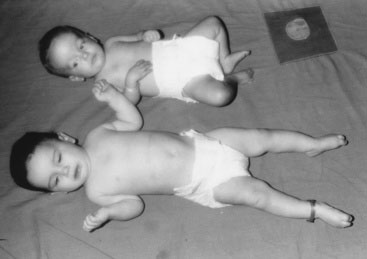
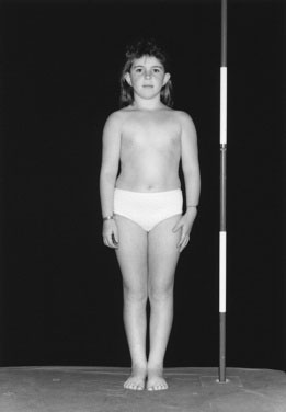
Children with GHD are described as cherubic (Figs. 16-2 through 16-4). They appear younger than their chronologic age, having a rounded face, chubby limbs, and “puddling” (or dimpling) of the anterior abdominal fat. Because of their infantile appearance, further enhanced by a high-pitched reedy voice, adults may baby them or classmates may carry them around like the class pet.

FIGURE 16–2 Hypoglycemia in infancy. The larger infant had hyperinsulinism in the neonatal period, which was treated by pancreatectomy. She is shown here at age 12 months beside a very small infant of the same age who has hypoglycemia secondary to GH deficiency.

FIGURE 16–3 Crystal, showing the typical features of GHD before treatment. Inset is the growth curve after treatment with GH.
Examination must include careful funduscopic and visual field assessment. You can see the poorly developed muscle bulk of a child with GHD on inspection and appreciate it by running your hands over the limbs. The hair is fine and wispy, and tooth eruption is delayed. In boys, the penis may be quite small (less than 2.5 cm); remember to push the suprapubic fat pad as flat as possible, in case a normal-sized penis is merely buried in a thick fat pad.
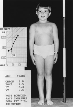
Height and weight percentiles (see Fig. 3–4) are usually discrepant in GHD, with a high weight-to-height ratio. This is a major clue to the need for detailed endocrine investigation. Chronic disease, especially inflammatory bowel disease, may be quite silent and manifest as short stature but, by contrast, with a low weight-to-height ratio.
When available, it is important to include the measurement of both biological parents of the child as a part of the assessment and to calculate the mid-parental height (see Chapter 3) to assess the child’s growth in the context of what would be expected for the family.
Investigate a growth velocity of less than 5 cm per year in children age 5 years to puberty; a growth velocity of less than 4 cm per year is clearly pathologic (see Chapter 3).
You cannot assess short stature or analyze growth velocity without examining the breasts and genitalia to determine the child’s pubertal stage (see Fig. 3–9).
Chief Characteristics of Thyroid Disorders in Children
Structure, function, signs, and symptoms
Typically, the history is the most important diagnostic aid in evaluating thyroid disorders, although the physical findings serve to bolster your suspicions. Thyroid size does not reflect thyroid function. First sort out the functional state: hypothyroid, hyperthyroid, or euthyroid (normal function). Then examine the thyroid to see whether it is too big or contains any lumps and to search for associated lymphadenopathy.
Evaluating a Child with Hyperthyroidism
Symptoms of hyperthyroidism are the same in children as in adults, but it is the parents or teachers who usually complain about the child’s restlessness and irritability. The child often does not complain, although the symptoms are obvious to everyone else. The following questions will elicit the essentials:
Evaluating a Child with Hypothyroidism
Like children with hyperthyroidism, children with hypothyroidism rarely complain of symptoms (Box 16–1) perhaps because the changes develop so insidiously that neither child nor parent notices the difference. When symptoms go unrecognized for years, linear growth is profoundly impaired and may be the presenting feature.
Causes of Childhood Hypothyroidism
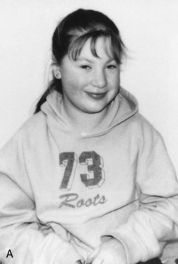
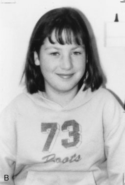
The most common cause of hypothyroidism in childhood is autoimmune thyroiditis (Hashimoto’s disease, or lymphocytic thyroiditis). Lymphocytic infiltration causes firm diffuse enlargement of the thyroid and a pebbled surface. The anterior pituitary, sensing the lack of thyroid hormone (loss of feedback inhibition), increases the secretion of thyroid-stimulating hormone (TSH), causing the thyroid to enlarge further. Although the gland can grow in response to TSH, it cannot necessarily keep up in terms of thyroxine (T4) production. Because the autoimmune process is destructive, some patients have a small or undetectable thyroid gland at presentation. A child with hypothyroidism secondary to hypothalamic-pituitary disease does not have thyroid enlargement (a goiter).
Congenital hypothyroidism is usually due to either absence of the thyroid or a hypoplastic thyroid that may be ectopic (located anywhere along the embryologic line of descent from the back of the tongue to the anterior neck.)
Case History 2
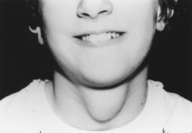
History. Shauna, age 15 years, says, “I was the tallest in my class in fifth grade, but I have not grown since then, and my periods haven’t started” (Fig. 16–5).


FIGURE 16–5 A, Shauna with the typical facial features of profound hypothyroidism. Note the puffy face and neck and the coarse, dry hair. B, Shauna after 4 months of treatment with thyroid hormone.
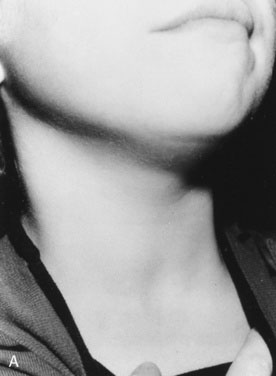
Your physical examination reveals all of the physical findings for hypothyroidism that are listed in Table 16–1. You particularly note the characteristic puffiness around Shauna’s upper face and eyes. In this case, because of profound hypothyroidism, there is also a more generalized swelling, including the neck.
TABLE 16–1 Causes of Precocious Puberty
Type of Precocious Puberty | Underlying Cause |
---|---|
Central | IdiopathicHypothalamic hamartomaGerminoma (pinealoma)Other cerebral tumors or malformationsSevere hypothyroidismSecondary to cranial irradiation |
Gonadal | Benign precocious thelarche (? central)Ovarian cysts, benignMcCune-Albright syndrome (polyostotic fibrous dysplasia of bone, ovarian cysts, large irregular café-au-lait spots)Leydig cell hyperplasia (familial testotoxicosis)Gonadal tumors (male and female)Isolated premature menses (? central) |
Adrenal | Benign premature adrenarcheCongenital adrenal hyperplasiaAdrenal tumors |
Iatrogenic | Exogenous sources |
With treatment for hypothyroidism, Shauna’s menses start almost immediately. She grows an inch and loses 4 kg (9 lb) during 4 months of therapy.
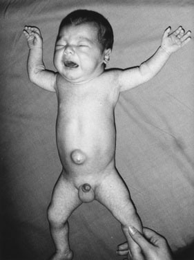
In a few infants, hypothyroidism is caused by a congenital enzyme defect in thyroid hormonogenesis. In such instances, a goiter is present. In many countries, congenital hypothyroidism is now detected in the presymptomatic stage, before any significant intellectual deficit occurs, through blood spot screening between the second and fifth days of life. If congenital hypothyroidism is left untreated, the clinical manifestations may not become fully apparent until around the third month of life.
Thyroid hormone is essential for adequate development of brain cell function and interneuronal connections. With absence of the hormone over the first few months of life, the infant suffers marked and irreversible developmental delay, the magnitude of the intellectual deficit increasing with each passing week. Universal newborn screening for hypothyroidism is therefore essential.
The infant with untreated congenital hypothyroidism has a low core body temperature, slow pulse rate, prolonged neonatal jaundice (because of poor hepatic conjugation of bilirubin), dry skin and hair, a large posterior fontanel (greater than 0.5 cm), poor head control, poor muscle tone, an enlarged tongue, a characteristically puffy face, and a hoarse cry (Fig. 16–6). The parents may report poor feeding and constipation. If acquired hypothyroidism develops after age 2 or 3 years, no permanent intellectual deficit is thought to occur.
Acquired hypothyroidism must be ruled out in any child with unexplained growth retardation. The child’s appetite is generally diminished, yet there is a modest weight gain, leading to a decrease in height-to-weight ratio. Such children are not massively obese; by contrast, prepubertal obese children generally grow well in height. Constipation is a common feature. In children with type 1 diabetes mel litus, you must always rule out the coexistence of autoimmune thyroid disease, usually lymphocytic thyroiditis, because these disorders are commonly related.
Approach to the Physical Examination of the Child with a Thyroid Disorder
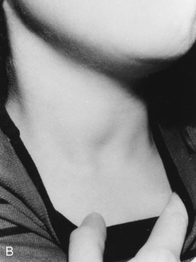
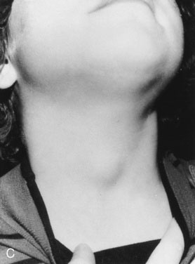
As with other pediatric examinations, always begin with the hands-off approach. Children are often ticklish, and touching the neck may elicit giggles and squirming, making it difficult to see the thyroid. Sit the child on a chair or examining table, with good light and in a relaxed atmosphere. If the child is old enough and cooperative, ask him or her to take a sip of water, first hold the water in the mouth without swallowing, and then swallow. Observe the area just above the sternal notch to see whether the small thyroid gland moves up and down during swallowing (Fig. 16–7). Next, ask the child to hyperextend the neck. This maneuver makes thyroid enlargement more obvious, and sometimes it is the only way to see the gland easily.


FIGURE 16–7 Demonstration of thyroid enlargement in a 12-year-old girl with a nodule in the right lobe. A, Little is visible at rest. B, The enlarged right lobe moves upward and becomes more obvious with swallowing. C, With neck extension, it is readily visible. D, Palpation of the nodule from the front.
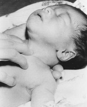
Examining newborns and young infants
In a newborn or young infant, use a different technique to examine the thyroid. Place your hand under the child’s back, between the scapulae, and raise the baby’s shoulders, allowing the head to fall back gently until it rests on the examining table or a parent’s lap. If the thyroid is enlarged, this maneuver will expose it, allowing you to palpate with the second and third fingers of your other hand (Fig. 16–8).
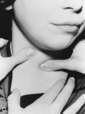
Examining young children and adolescents
In the young child or adolescent, first observe the gland. Then, after gaining the child’s cooperation, palpate it. We generally examine from the front, as follows. Locate the cricoid cartilage, which is the smaller tracheal cartilage, just below the notched thyroid cartilage or Adam’s apple. Palpate just below the cricoid cartilage for a soft ribbon of tissue that moves with swallowing and is the normal isthmus. With practice you will become expert at finding the thyroid isthmus. Then rest both thumbs gently over the lobes of the thyroid, and ask the child to swallow. While the child swallows, move your thumbs with the child’s skin, up and down over the thyroid to outline its size. Note any nodules or irregularities you feel. You can also examine the thyroid in the traditional way, from behind the child: Place the second and third fingers of your examining hand over the gland to estimate its size, and ask the child to swallow. We often joke with older children who hate to have their necks touched that we have never choked anyone and are not allowed to!
Always measure and record the gland size so that any significant changes between visits are documented. With a measuring tape, measure from upper to lower pole and the greatest vertical dimension of the isthmus. Record the results in diagrammatic form, indicating any nodules or other irregularities.
The normal thyroid weight in grams corresponds approximately to the child’s age in years; a normal adult gland weighs 20 to 25 g and is the size of your thumb from the distal interphalangeal joint to the tip. The thyroid can be palpated in most older children. Because its normal consistency is soft, the contours can barely be seen, and the gland is rather flat when the child hyperextends the neck. Once the thyroid gland becomes firm, as in autoimmune thyroiditis (Hashimoto’s disease), or hard, as in a carcinoma, you should recognize the difference in consistency, even though enlargement may not be striking. Some carcinomas, however, may not be hard to the touch because consistency is not always a reliable diagnostic sign.
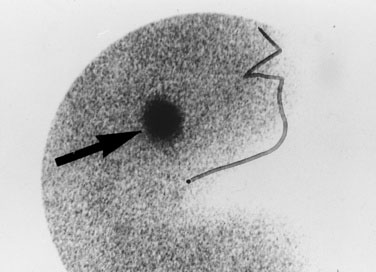
Most midline neck masses are thyroid glands or thyroid remnants. A central rounded midline mass between the thyroid and the chin is almost certain to be a thyroglossal duct cyst (Fig. 16–9), derived from a remnant of the thyroglossal duct, which runs from the foramen cecum of the tongue downward to the cricoid cartilage during fetal development. When the child sticks out the tongue, the mass moves upward. The child may present initially with an infected thyroglossal duct cyst. Sometimes, the gland fails entirely to migrate downward from the foramen cecum and manifests clinically as an oval or rounded midline mass at the base of the tongue (Fig. 16–10). This lingual thyroid usually represents all of the thyroid that the child has. Most lingual thyroids are treated with daily therapy using l-thyroxine, which suppresses pituitary thyrotropin (TSH) secretion, causing the lump to shrink markedly.

FIGURE 16–10 Thyroid scan in an infant demonstrating uptake of the radionuclide at the base of the tongue in a lingual thyroid (arrow).
The thyroid may be absent, small, ectopic, or enlarged. If both lobes are enlarged fairly symmetrically, it is described as diffusely enlarged. The normal gland may have slight undulations on palpation, but if you find one or more distinct lumps, the child has either a solitary nodule or a multinodular goiter.
The uninformative term goiter denotes any thyroid enlargement but reveals nothing about the functional or anatomic diagnosis. A diffuse or nodular enlargement may give the child a sensation of difficulty swallowing but rarely causes true dysphagia. Press your finger gently over your own trachea just above the sternal notch to understand the sensation that children feel with even slight thyroid swelling. Enlargements may be painful and tender to touch. A change in the voice may occur if the enlargement damages the recurrent laryngeal nerve or if the vocal cords become myxedematous in severe hypothyroidism.
A diffusely enlarged, nontender gland that is soft to somewhat firm in consistency is compatible with Graves disease. By contrast, a tender, diffusely swollen gland in a child who withdraws when the gland is touched is likely to represent subacute thyroiditis attributed to a viral cause. Thyroid-stimulating antibodies interact with the TSH receptor on the thyroid cell, producing the constant stimulation of Graves disease. In subacute thyroiditis, disrupted cells produce a sudden thyroid hormone discharge, causing a transient hyperthyroid period, which is often followed by a longer hypothyroid state, then by euthyroidism within 3 to 6 months. A similar sequence is seen in a condition called silent thyroiditis, so called because it is not painful. Silent thyroiditis is most common in the postpartum period but does occur at all ages and in both sexes; it has an autoimmune etiology.
If one or more nodules are found in a child with hyperthyroidism, the cause is probably a benign adenoma, called toxic nodular goiter, which is overproducing thyroid hormone. This condition is seen more commonly in adults. The discrete smooth, oval or round nodules are firmer than the surrounding gland but not rock-hard. When the child swallows, they move up and down and are not fixed to extrathyroidal tissues. Rare TSH-secreting pituitary adenomas can cause diffuse thyroid enlargement and systemic signs of hyperthyroidism but without the eye findings characteristic of autoimmune (Graves) disease.
An eye examination yields important clues for diagnosis and management of thyroid disease. In hyperthyroidism, the palpebral fissures are widened and blinking is diminished, giving the child a staring appearance. In autoimmune Graves disease, the eye muscles can swell to several times their normal size, and the retro-orbital fat pad enlarges, causing the eye to bulge out of the orbit (proptosis), often with injection (prominent small surface vessels) and edema of the bulbar conjunctiva (chemosis). Because individuals with proptosis have difficulty closing their lids, corneal ulceration may occur, causing red, sore eyes, particularly in the morning. Ask the parent whether the child closes the eyes when sleeping. Without adequate tears and with open lids, the cornea becomes dry and vulnerable to ulceration. Enlarged ocular muscles produce an imbalance of extraocular muscle function, sometimes sufficient to cause diplopia, which may be unrecognized by affected patients until they are asked to look upward and outward, the direction of gaze in which diplopia can first be elicited. Usually, there is some loss of convergence, and the child cannot cross the eyes to the usual extent when trying to follow the physician’s finger as it is moved toward the nose.
Thyroid bruits are characteristic of Graves disease, but this sign is not very useful in children. The problem with auscultating the neck in children is that if you press hard enough with the stethoscope, you can make anything whistle.
Infants may be born with neonatal hyperthyroidism from transplacental passage of thyroid-stimulating immunoglobulin, an antibody produced in a mother who has active or treated Graves disease with persistent circulating antibody. Although these babies have the same signs as older children, they are more likely to have hyperthermia and cardiac tachyarrhythmias and to develop congestive heart failure.
Neonatal hyperthyroidism must be treated, but it is a self-limited condition. It subsides when maternal immunoglobulin G (IgG) disappears from the infant’s circulation.

Stay updated, free articles. Join our Telegram channel

Full access? Get Clinical Tree

