Chapter 35 Climacteric
MENOPAUSE AND PERI- AND POSTMENOPAUSE∗
As average life expectancy increases in the United States and elsewhere, women and men are often living well into their ninth decade of life. The preservation of their quality of life in terms of both physical and mental activity is a high priority for them. Many women will live for 30 to 40 years after reproductive function ends.
The climacteric refers to the phase in a woman’s reproductive life when a gradual decline in ovarian function results in decreased sex steroid production with its sequelae. Because this phase is a normal consequence of the aging process, it should not be considered an endocrinopathy.
Menopause literally refers to the last menstrual period. The exact time of menopause is usually determined in retrospect; that is, 1 year without menses. In most women, menopause occurs between the ages of 50 and 55 years, with an average age of 51.5 years, but some have their menopause before the age of 40 (premature menopause), whereas a few may menstruate until they are in their 60s.
Women are born with about 1.5 million oocytes (primary ovarian follicles) and reach menarche (first menstruation) with about 400,000 potentially responsive eggs. Most women ovulate about 400 times between menarche and menopause, and during this time, nearly all other oocytes are lost through atresia. When the oocytes either have ovulated or become atretic, the ovary becomes minimally responsive to pituitary gonadotropins, the ovarian production of estrogen and progesterone ends, and ovarian androgen production is reduced. These hormone alterations often result in unpleasant and even harmful physical, psychological, and sexual changes in postmenopausal women and can have a negative impact on their quality of life.
Hormonal Changes
Menopause rarely occurs as a sudden loss of ovarian function. For some years before menopause, the ovary begins to show signs of impending failure. Anovulation becomes common, with resulting unopposed estrogen production and irregular menstrual cycles (see Chapter 33). On occasion, heavy menses, endometrial hyperplasia, and increasing mood and emotional changes may occur. In some women, hot flashes (or flushes) and night sweats begin well before menopause is reached. These perimenopausal symptoms may last 3 to 5 years before there is complete loss of menses and postmenopausal levels of hormones are reached.
Some women continue to produce estrogen indirectly in substantial amounts for many years after menopause. Androstenedione from the ovary and the adrenal gland is converted in peripheral fat tissues to estrone, which is then capable of maintaining the vagina, skin, and bone in reasonable cellular tone and reducing the incidence of flashes. Although this unopposed estrogen may be beneficial to women, it may also be responsible for the increased incidence of endometrial or breast cancer, particularly among obese women. For this reason, it is important that postmenopausal women have regular breast examinations and, if abnormal vaginal bleeding occurs, endometrial sampling.
Ovarian Senescence
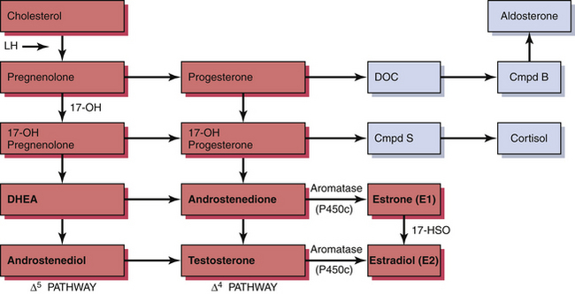
The ovary produces a sequence of hormones during a normal menstrual cycle. Under the influence of luteinizing hormone (LH), cholesterol from the liver is used to produce the androgens androstenedione and testosterone in the theca cells of the ovarian follicle. They, in turn, are converted in the granulosa cells immediately surrounding the oocytes into estrogen. Following ovulation, the luteal cells (luteinized granulosa cells) manufacture and secrete progesterone as well as estrogen. The synthesis of these sex hormones depends on the presence of viable follicles and ovarian stroma and the production of follicle-stimulating hormone (FSH) and LH in adequate amounts to induce their biosynthetic activity. The ovarian and adrenal (for comparison) steroid biosynthetic pathways are depicted in Figure 35-1.

FIGURE 35-1 Diagrammatic representation of the steroid biosynthetic pathways. Ovarian sex steroid pathways are in red and adrenal in blue. Cmpd B, corticosterone; Cmpd S, II-deoxycortisol; DH, hydroxylase; DHEA, dehydroepiandrosterone; DOC, desoxycorticosterone; LH, luteinizing hormone; P450c, cytochrome P450.
ESTROGEN
Following menopause, estradiol (E2) values decline (to only 10 to 50 pg/mL), but estrone levels may increase. Estrone (E1) can be produced by peripheral conversion of androstenedione from the ovary and the adrenal gland. In some women, the amount of postmenopausal estrogen may be considerable.
ANDROGENS
Women normally produce significant quantities of androgens by the metabolic conversion of cholesterol to both androstenedione and testosterone. Although the major portion of androgen is aromatized to estrogen, some androgen circulates. After menopause, there is a decrease in the level of circulating androgens, with androstenedione falling to less than half that found in normal menstruating young women, whereas testosterone gradually diminishes over about 3 to 4 years. Even though postmenopausal women produce less androgens, they tend to be more sensitive to them because of the lost opposition of estrogen. This sometimes results in unwelcome changes such as excessive facial hair growth and decreased breast size.
PROGESTERONE
With anovulation during the climacteric and ovarian failure after the menopause, the production of progesterone declines to low levels. The minimal progesterone present is insufficient to induce those cytoplasmic enzymes (estradiol dehydrogenase and estrone sulfuryltransferase) that convert estradiol to the less potent estrone sulfate and to reduce the levels of cellular estrogen receptors. Altogether, this may result in increased estrogen-induced mitosis in the endometrium. The absence of progesterone also prevents the secretory histologic transformation in the endometrium and its subsequent sloughing. As a consequence, perimenopause is often associated with irregular vaginal bleeding, endometrial hyperplasia and cellular atypia, and an increased incidence of endometrial cancer.
GONADOTROPINS
The two gonadotropins, LH and FSH, are produced in the anterior pituitary gland. When levels of estrogen are low, the arcuate nucleus and paraventricular nucleus in the hypothalamus are freed from negative feedback and are able to secrete increasing amounts of gonadotropin-releasing hormone (GnRH) into the pituitary portal circulation. This, in turn, stimulates an increased release of LH and FSH into the circulation. The higher central nervous system neurotransmissions responsible for the increased pulsatile release of GnRH (and subsequent gonadotropin release) are also thought to have parallel effects elsewhere in the hypothalamus, especially the body temperature control region. This leads to sudden induction of increased skin blood flow and perspiration—the hot flash that so characterizes menopause. Typical levels of FSH in postmenopausal women are greater than 20 or 40 IU/L, depending on the assay used.
Clinical Manifestations
Loss of estrogen is associated with urogenital atrophy and osteoporosis (Table 35-1). Although postmenopausal women have a higher incidence of heart disease and of cancer, the relationship between these adverse events and reduced endogenous estrogen production, as well as the effects of hormonal therapy on them, remains unclear and controversial.
TABLE 35-1 CONSEQUENCE OF ESTROGEN LOSS
Symptoms (early) | |
Physical changes (intermediate) | |
Diseases (late) |
![]() Stay updated, free articles. Join our Telegram channel![]() Full access? Get Clinical Tree![]() ![]() ![]() |