118 Chromosomal Abnormalities
Since the discovery in 1959 that Down syndrome is caused by trisomy of chromosome 21, multiple syndromes have been found to have an identifiable chromosomal abnormality. These range from triplication of a complete chromosome as in Edwards syndrome (trisomy 18) to monosomies such as Turner syndrome (45,X) and to a growing number of smaller duplication and deletion syndromes. Several of the more common or illustrative chromosomal aberrations are discussed in this chapter.
Trisomy 18 (Edwards Syndrome)
Etiology and Pathogenesis
Trisomy 18 (Edwards syndrome) is most commonly caused by meiotic nondisjunction, and the risk is related to maternal age. However, Edwards syndrome can also result from an unbalanced translocation. If the parents of an affected infant have a normal karyotype (i.e., neither is a balanced translocation carriers), the recurrence risk for trisomy 18 in a subsequent pregnancy is no greater than the age-related risk.
Clinical Presentation
Trisomy 18 is one of the more common malformation syndromes, with an incidence of 0.3 in 100 newborn infants. On prenatal ultrasound, increased nuchal translucency may be seen early in gestation. At birth, reduced birth weight, a small placenta, and fetal distress may be noted along with multiple other clinical features (Table 118-1).
Table 118-1 Frequent Clinical Features in Trisomies 18 and 13
ASD, atrial septal defect; CNS, central nervous system; PDA, patent ductus arteriosus; VSD, ventricular septal defect.
Prognosis
The median survival of infants with trisomy 18 is 2 weeks, with 5% to 10% of patients living through the first year. Surviving children exhibit severe physical and mental retardation with vision and hearing impairment. Poor feeding often makes nasogastric tube feedings necessary. Growth is poor, and specific growth charts are available.
Trisomy 13 (Patau Syndrome)
Etiology and Pathogenesis
Most cases of Patau syndrome are caused by trisomy 13, which also demonstrates an increased incidence with advancing maternal age. Similarly, chromosomal studies should be performed to rule out an unbalanced translocation because recurrence is higher in a parent with a balanced translocation.
Clinical Presentation
Trisomy 13 occurs with an incidence of approximately 1 in 5000 births. Early prenatal detection is possible with a combined screening protocol that includes maternal age, nuchal translucency, and maternal serum markers. The mean birth weight is reduced, and multiple dysmorphic features may be observed (see Table 118-1).
Turner’s Syndrome (45,X)
Etiology and Pathogenesis
Turner’s syndrome is diagnosed in a female by the absence of a second normal sex chromosome caused by the complete or partial deletion of an X or Y chromosome. The loss is not associated with advanced maternal age and may happen during spermatogenesis, oogenesis, or postzygotic nondisjunction. The resulting karyotype is 45,X in approximately half of patients with Turner syndrome, but isochromosomes with duplication of the long arm of the X chromosome, ring and marker chromosomes, and mosaicism are frequently observed.
Turner syndrome occurs in 1 in 2500 to 1 in 3000 female live births and is caused by the absence of a second normal sex chromosome. The incidence of 45,X is actually higher in pregnancy losses.
Clinical Presentation
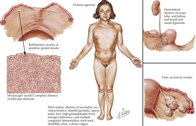
Females with Turner syndrome may present prenatally with features of lymphedema/hydrops and/or a congenital heart defect, in puberty due to short stature or in adulthood with premature ovarian failure (Figure 118-1). Multiple organ systems may be involved, but no single feature is pathognomonic or universally present.
Several physical features are found as a consequence of lymphatic abnormalities that include webbed neck, puffy hands and feet, dysplastic finger- and toenails, protuberant ears, and a broad chest.
Up to 45% of patients have a cardiac anomaly identified by echocardiography, most often a bicuspid aortic valve or aortic coarctation, but other cardiac anomalies such as partial anomalous pulmonary venous connection can also occur. Renal anomalies are equally frequent (40%), including renal agenesis and horseshoe kidney. Hypothyroidism may develop in 5% to 10% of girls in mid to late childhood and affects up to one-third of adult women.
Growth delay begins in childhood, but short stature may not be noticed until 5 to 10 years of age or in puberty with a blunted growth spurt. Growth hormone treatment may lead to improvement, but adult height is usually reduced.
Gonadal dysgenesis is a hallmark of Turner syndrome and typically leads to ovarian failure. Puberty occurs spontaneously in 10% of patients, and hormonal replacement therapy is necessary to achieve development of secondary sexual characteristics as well as to avoid osteoporosis and other postmenopausal complications. Nonetheless, spontaneous pregnancy has been observed in about 2% to 4% of women with Turner syndrome.
Most girls with Turner syndrome have normal developmental milestones and intelligence. About 10% of individuals experience specific learning disabilities, mental retardation, motor deficits, or difficulties with attention, maturity, and social skills. Correlation has been observed between the intellectual phenotype and the karyotype, and girls with a marker or ring chromosome have a higher prevalence of mental retardation.
Klinefelter Syndrome
Etiology and Pathogenesis
Klinefelter syndrome is caused by a 47,XXY or 46,XY/47,XXY karyotype with mosaic men exhibiting a milder phenotype. Nondisjunction of the sex chromosomes is equally derived from either parent with only a small association with maternal age.
With an incidence of 1 in 500 to 1 in 1000 male infant births, Klinefelter syndrome is the most common sex chromosome aneuploidy.
Clinical Presentation
Newborns with 47,XXY do not have any significant dysmorphic features. In fact, 10% of boys are identified incidentally during chorionic villus sampling or amniocentesis for advanced maternal age. In rare cases, chromosomal studies are initiated in the newborn period for cryptorchidism, hypospadias, or a small phallus. About 25% of affected individuals are diagnosed in childhood, adolescence, and adulthood. A large fraction of 47,XXY men are thought to remain undiagnosed.
In childhood, language delay, learning differences, or behavioral problems may bring a boy to medical attention. Physical symptoms start to develop in adolescence with increased height, narrower than average shoulders, broader than average hips, and scoliosis or kyphosis.
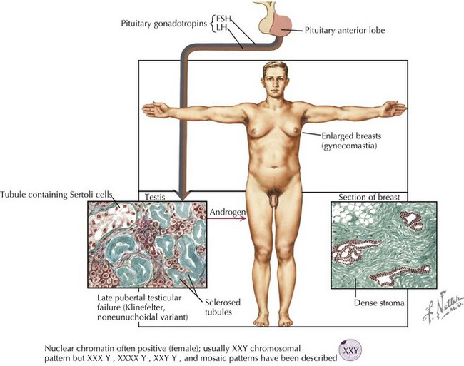
Although the age and onset of puberty is unchanged in boys with Klinefelter syndrome, testicular growth is arrested, leading to oligospermia or azoospermia and low testosterone levels. A small fraction of men present with gynecomastia, eunuchoid habitus, and decreased hair (Figure 118-2). Muscle strength may be decreased. Testosterone replacement therapy aims at increasing muscle mass, libido, energy, and improving mood through support of androgen-dependent processes. Fertility is rarely preserved except in mosaic men.

Stay updated, free articles. Join our Telegram channel

Full access? Get Clinical Tree

