Chapter 38 Cervical Dysplasia and Cancer
Cervical cancer kills about 250,000 women a year worldwide and is the most common cause of death from cancer in women. About 80% of new cases reported each year occur in developing countries. In developed countries, regular screening with Papanicolaou (Pap) smears has markedly decreased the incidence of the disease, and most cases now occur in women who have not had regular Pap smears. In the United States, cervical cancer now ranks only 13th among cancers in women, with 11,150 new cases expected in 2007, and 3,670 deaths.
Studies have identified persistent infection with a high-risk human papillomavirus (HPV) as the cause of virtually all cervical cancers. Recent randomized clinical trials of prophylactic HPV vaccines have demonstrated dramatic efficiency in preventing HPV 16 and 18 infections as well as precancerous cervical lesions. Although it will take several decades to demonstrate a decreased incidence of invasive cervical cancer, with widespread use, HPV vaccination has the potential to markedly decrease the incidence of cervical cancer in future generations.
Etiology and Epidemiology
There are 15 high-risk HPV types, and types 16 and 18 are responsible for 70% of cervical cancers. Types 6 and 11 have been associated with cervical condylomas and low-grade cervical intraepithelial neoplasia (CIN).
The adolescent cervix is believed to be more susceptible to carcinogenic stimuli because of the active process of squamous metaplasia, which occurs within the transformation zone during periods of endocrine change. This squamous metaplasia is normally a physiologic process, but under the influence of the HPV, cellular alterations occur that result in an atypical transformation zone. These atypical changes initiate CIN, which is the preinvasive phase of cervical cancer.
Cervical cancer and its precursors have been associated with several epidemiologic variables (Box 38-1). These risk factors basically increase the likelihood of exposure to a high-risk HPV type.
The disease is relatively rare before 20 years of age, and the mean age is about 47 years.
Primary Prevention
Two prophylactic vaccines are presently available. The quadrivalent vaccine Gardasil, which is manufactured by Merck (Whitehouse Station, NJ) and protects against HPV types 6, 11, 16, and 18, was approved by the U.S. Food and Drug Administration in June 2006 for females aged 9 to 26 years. The bivalent vaccine Cervarix, which is manufactured by GlaxoSmithKline (Philadelphia, Pa) and protects against HPV types 16 and 18, was approved by the Australian Therapeutic Goods Administration in April 2007 for use in females aged 9 to 45 years.
HPV vaccination is most effective if performed before the onset of sexual activity. Vaccination is still recommended after commencement of sexual activity, and even after prior abnormal cytology or CIN, but it is likely to be less effective after HPV exposure. Australia was the first country in the world to introduce HPV vaccination into the National Immunization Program. In 2007, vaccination with Gardasil was introduced for all schoolgirls aged 12 years.
Screening of Asymptomatic Women
The American College of Obstetricians and Gynecologists has recommended that all women undergo an annual physical examination, including a Pap smear, within 3 years of sexual intercourse, or by age 21 years. Annual screening should occur until age 30 years. If there have been three consecutive negative tests, screening may occur every 2 or 3 years at the discretion of the treating physician. Both the endocervical canal and the ectocervix should be sampled when taking the Pap smear.
The false-negative rate for conventional Pap smears for high-grade intraepithelial lesions is generally reported to be about 20%, but it is higher for glandular lesions and for invasive cancers.
New technologies have been developed to decrease the false-negative rate. ThinPrep (Cytyc, Marlborough, Mass) and SurePath (BD Diagnostics–TriPath, Franklin Lakes, NJ) are automated liquid-based slide-preparation systems. With liquid-based cytology, the spatula or brush taking the smear is placed into a fixative solution, instead of smearing the cells directly onto a glass slide. Blood, mucus, and inflammatory cells are eliminated, and a monolayer smear is then automatically prepared by a machine. BD Focal Point (BD Diagnostics–TriPath) and ThinPrep Imager (Cytyc) are computerized image processors that select the most abnormal cells on a slide. They increase the sensitivity of slide reading, while decreasing the time needed by the cytotechnician to read each slide, thereby improving the cost-effectiveness of screening.
The cost-effectiveness of HPV DNA testing as a primary screening test, either alone or in combination with cervical cytology, in women aged 30 years or older, is currently under investigation. HPV DNA testing is much more sensitive than cervical cytology, but less specific.
Women should have regular cervical screening even if they have received the HPV vaccine because the vaccine does not protect against all high-risk HPV viral types.
Cervical Topography
During early embryonic development, the cervix and upper vagina are covered with columnar epithelium. During intrauterine development, the columnar epithelium of the vagina is progressively replaced by squamous epithelium. At birth, the vagina is usually covered with squamous epithelium, and the columnar epithelium is limited to the endocervix and the central portion of the ectocervix. In about 4% of normal female infants and about 30% of those exposed to diethylstilbestrol in utero, the columnar epithelium extends onto the vaginal fornices. Macroscopically, the columnar epithelium has a red appearance because it is only a single cell layer thick, allowing blood vessels in the underlying stroma to show through it.
The embryologic squamous and columnar epithelia are designated the original and native squamous and columnar epithelia, respectively. The junction between them on the ectocervix is called the original squamocolumnar junction.
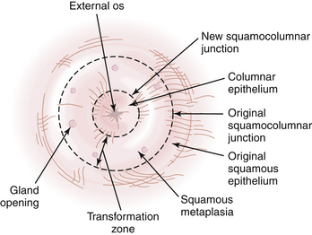
Throughout life, but particularly during adolescence and a woman’s first pregnancy, metaplastic squamous epithelium covers the columnar epithelium so that a new squamocolumnar junction is formed more proximally. This junction moves progressively closer to the external os and then up the endocervical canal. The transformation zone is the area of metaplastic squamous epithelium located between the original squamocolumnar junction and the new squamocolumnar junction (Figure 38-1).
Classification of an Abnormal Papanicolaou Smear
In 1988, a consensus meeting was convened by the Division of Cancer Control of the National Cancer Institute to review existing terminology and to recommend effective methods of cytologic reporting. As a result of this meeting, the Bethesda system was devised and requires (1) a statement regarding the adequacy of the specimen for diagnosis, (2) a diagnostic categorization (normal or other), and (3) a descriptive diagnosis. A revised Bethesda system was developed in 2001 and is shown in Box 38-2.
BOX 38-2 Bethesda Classification of Cytologic Abnormalities (2001, Abridged)
Epithelial Cell Abnormalities
Squamous Cell
CERVICAL INTRAEPITHELIAL NEOPLASIA
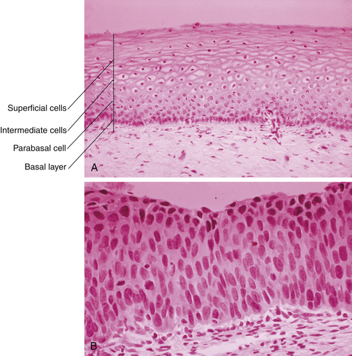
CIN represents a spectrum of disease, ranging from CIN I (mild dysplasia) to CIN III (severe dysplasia and carcinoma in situ). At least 35% of patients with CIN III develop invasive cancer within 10 years, whereas lower grades of CIN often spontaneously regress. With CIN, there is abnormal epithelial proliferation and maturation above the basement membrane. Involvement of the inner one third of the epithelium represents CIN I, involvement of the inner one half to two thirds represents CIN II, and full-thickness involvement represents CIN III (Figure 38-2). The disease is asymptomatic.

FIGURE 38-2 Histologic appearance of normal cervical squamous epithelium (A) and carcinoma in situ (B) of the cervix. In the normal epithelium, note the orderly maturation from the basal layer to the parabasal cells, glycogenated intermediate cells, and flattened superficial cells. In the carcinoma in situ, the entire thickness of the epithelium is replaced by immature cells that are variable in size and shape and have irregular nuclei. Mitotic figures are seen in the lower two thirds of the epithelium.
COLPOSCOPY
The colposcope is a stereoscopic binocular microscope of low magnification, usually 10× to 40×. Illumination is centered, and the focal length is between 12 and 15 cm.
To perform a colposcopic examination, an appropriately sized speculum is inserted to expose the cervix, which is cleansed with a cotton pledget soaked in 3% acetic acid to remove adherent mucus and cellular debris. A green filter can be employed to accentuate the vascular changes that frequently accompany pathologic alterations of the cervix.
At colposcopy, the original or native squamous epithelium appears gray and homogeneous. The columnar epithelium appears red and grape-like. The transformation zone can be identified by the presence of gland openings that are not covered by the squamous metaplasia and by the paler color of the metaplastic epithelium compared with the original squamous epithelium. Nabothian follicles may also be seen in the transformation zone. Normal blood vessels branch like a tree.
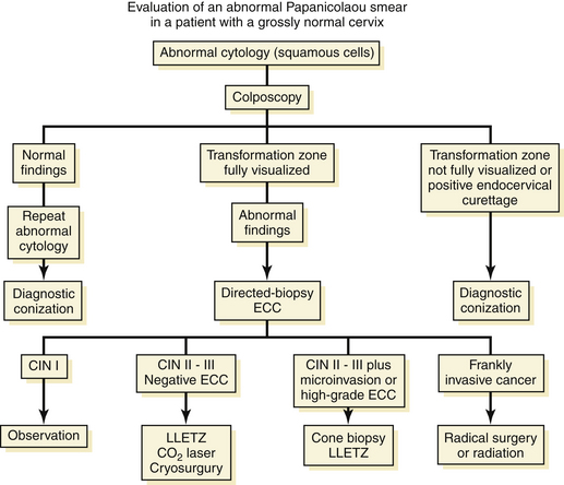
Evaluation of a Patient with an Abnormal Papanicolaou Smear
An algorithm for the evaluation of patients with abnormal Pap smears is presented in Figure 38-3.

FIGURE 38-3 Algorithm for evaluation of patients with an abnormal Papanicolaou smear and a grossly normal-appearing cervix. CIN, cervical intraepithelial neoplasia; ECC, endocervical curettage; LLETZ, large loop excision of the transformation zone.
Any patient with a grossly abnormal cervix should have a punch biopsy performed, regardless of the results of the Pap smear.

Stay updated, free articles. Join our Telegram channel

Full access? Get Clinical Tree

