CEREBROSPINAL FLUID EVALUATION

10 CEREBROSPINAL FLUID EVALUATION


General Discussion


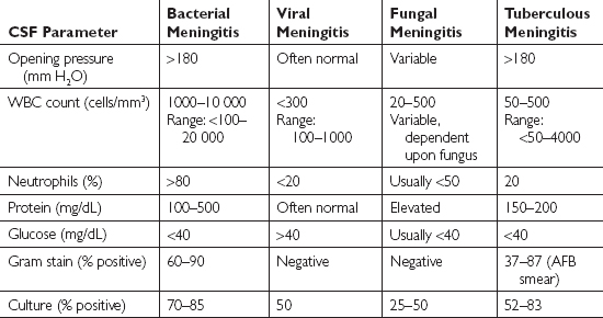
The central nervous system (CNS) is susceptible to bacterial, viral, and fungal infections, as well as to prion diseases and numerous local and systemic diseases. Examination of the cerebrospinal fluid (CSF) is crucial in helping to diagnose infections and other diseases. Although not definitive, certain CSF findings are suggestive of bacterial, viral, fungal, or tuberculous meningitis. These findings are outlined in Table 10.1 below.


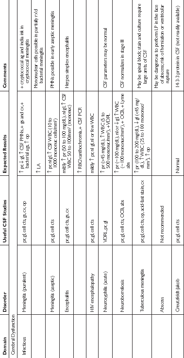
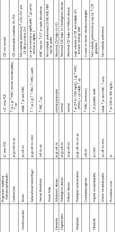
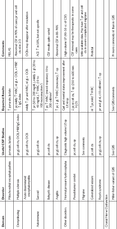
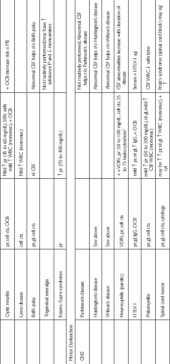
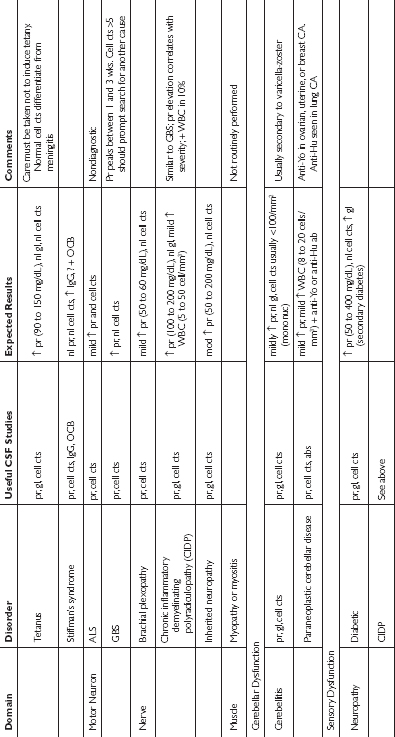
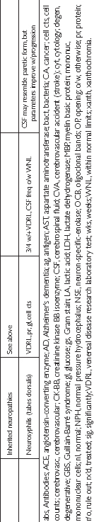
These typical findings, in combination with specific antigen, antibody, and polymerase chain reaction tests may help to reveal the origin of a CNS infection or disease. The tests that are ordered on the CSF should be guided by the suspected underlying cause of the patient’s illness. Recommended CSF studies based upon specific disorders are outlined in Table 10.2 below.


In patients who have bacterial meningitis and who receive antibiotics before lumbar puncture is performed, CSF abnormalities such as elevated white blood cell (WBC) count, elevated protein concentration, and depressed glucose may persist for one to three days, while results of Gram stain and culture of the CSF can become negative within hours after the antibiotics are administered. A mononuclear pleocytosis is usually present in patients who have viral meningitis, but it can be preceded by a transient predominance of polymorphonucleocytes for 8 to 48 hours. Elevated levels of CSF adenosine deaminase have a high sensitivity and specificity for tuberculous meningitis in adults.


For bacterial meningitis, CSF Gram stain and culture are the diagnostic tests of choice. Blood culture may also help identify the causative organism. CSF viral culture is able to detect 14 to 24% of cases of viral meningitis. Tuberculous and fungal meningitis may be difficult to diagnose by routine CSF smear or culture. CSF culture is positive in 52 to 83% of cases of tuberculous meningitis.


Cerebrospinal fluid cell counts with differentials should be performed on every specimen. Typically, the CSF contains no red blood cells (RBC)/μL and 0 to 1 WBC/μL. A traumatic lumbar puncture causes elevations of RBCs and WBCs, but these elevations are differentiated from subarachnoid hemorrhage because in a traumatic lumbar puncture the elevations are high in the first tube but clear in the later tubes. In subarachnoid hemorrhage, the elevations persist in each test tube. To determine whether an elevated CSF WBC is due to blood from a traumatic tap or other causes, an expected ratio may be used. If the elevated WBC is due to blood in the CSF, 1 WBC/μL for every 700 RBC/μL is found. If the WBC exceeds this ratio, its origin must be accounted for from other etiologies such as infection or inflammation.


The CSF glucose concentration is normally 60% of the plasma glucose concentration. It is important to obtain a serum glucose level at the time of the CSF sample. An elevated CSF glucose level results from an elevated plasma glucose level. A decreased CSF glucose concentration may be due to hypoglycemia, bacterial meningitis, fungal meningitis, certain viral meningitides, subarachnoid hemorrhage, carcinomatosis meningitis, chemical meningitis, and parasitic meningitis.


Elevation in CSF protein is a nonspecific but sensitive indicator of CNS disease. A CSF protein concentration greater than 500 mg/dL is an infrequent finding, but can occur with bacterial meningitis, subarachnoid hemorrhage, or spinal–subarachnoid block. When a significant amount of blood is present in the CSF, the total protein concentration can be corrected by reducing the protein by 1 mg/dL for every 1000 RBCs in the CSF. Protein concentrations of 100 mg/dL or greater have sensitivity and specificity for bacterial meningitis of 82 and 98%, respectively. If the concentration is 200 mg/dL, the sensitivity is 86% and the specificity is 100%.


The finding of oligoclonal bands in the CSF implies that a single clonal population of plasma cells is responsible for each band seen on gel electrophoresis. More than one oligoclonal band rarely occurs in normal CSF. A serum sample should be obtained simultaneously with a CSF sample to determine whether oligoclonal bands are unique to the CSF. Oligoclonal bands are present in 83 to 94% of patients with multiple sclerosis, and are also present in disorders such as subacute sclerosing panencephalitis, CNS lupus, neurosarcoidosis, cysticercosis, Behçet’s syndrome, Guillain-Barré syndrome, some brain tumors, and viral, fungal, and bacterial infections.

Stay updated, free articles. Join our Telegram channel

Aug 17, 2016 | Posted by in PEDIATRICS | Comments Off on CEREBROSPINAL FLUID EVALUATION

Full access? Get Clinical Tree

Get Clinical Tree app for offline access