CHAPTER 13 CARDIOVASCULAR PATHOLOGY
INTRODUCTION

DIAGNOSTIC ENDOMYOCARDIAL BIOPSY

CARDIOMYOPATHIES
Table 13.1 Simplified classification of pediatric cardiomyopathies (Richardson et al 1996)
Dilated cardiomyopathy |
Hypertrophic cardiomyopathy |
Restrictive cardiomyopathy |
Arrhythmogenic right ventricular cardiomyopathy |
Histopathological features of cardiomyopathies on biopsy
Dilated cardiomyopathy (Figs 13.1–13.4)

Fig 13.1 Photomicrograph of myocardium of a 2-year-old girl with dilated cardiomyopathy. The myofibers are elongated, stretched and wavy. The nuclei show mild enlargement and hyperchromasia.

Fig 13.2 Photomicrograph of an endomyocardial biopsy from a girl aged 11 years. There is prominent interstitial fibrosis. The myocytes show contraction bands. (Masson-trichrome)

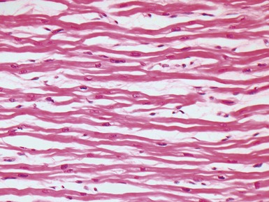
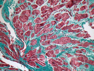
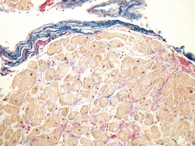
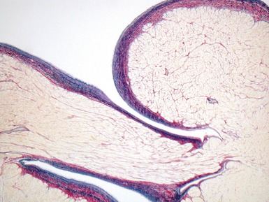
Fig 13.3 Photomicrograph of an endomyocardial biopsy from the same case as Fig 13.2. The myocytes show variation in size, as do their nuclei. There is mild interstitial fibrosis and the endocardium shows mild to moderate, fibroelastic thickening. (Elastic van Gieson)

Fig 13.4 Photomicrograph of myocardium from a 5-year-old boy with dilated cardiomyopathy. Scattered mast cells are evident in a perivascular location. While special staining may be used to demonstrate mast cells, they are usually readily apparent at high-power on H&E staining. Mast cell numbers are increased in the myocardium in dilated cardiomyopathy and in cases of myocarditis. (H&E)
Hypertrophic cardiomyopathy (Figs 13.5, 13.6)

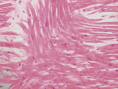
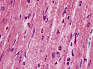
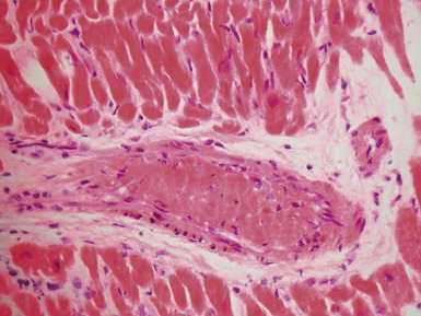
Fig 13.5 Photomicrograph of heart from an 11-year-old boy with hypertrophic cardiomyopathy. There is readily apparent myocyte disarray. Disarray is also usually evident at a macroscopic level where the muscle bundles are whorled, and also at the ultrastructural level where the myofilaments are disorganized.

Fig 13.6 Photomicrograph of a 7-month-old girl with hypertrophic cardiomyopathy due to mitochondrial respiratory chain complex-1 deficiency. Giant mitochondria are visible within the myocyte cytoplasm as rounded homogenous eosinophilic inclusions. (H&E)
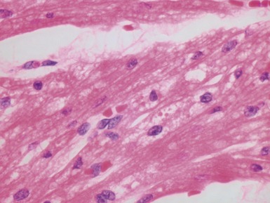
Table 13.2 Associations of myocyte disarray
Familial hypertrophic cardiomyopathy |
Restrictive cardiomyopathy |
Normal heart near the insertion of the septum into the ventricular free walls |
Congenital heart disease, particularly hypoplastic left heart |
Previous biopsy site on endomyocardial biopsy (see section on post-transplant biopsy) |
Adjacent to myocardial scars |
Restrictive cardiomyopathy
Arrhythmogenic right ventricular cardiomyopathy (Fig 13.7)

Fig 13.7 Photomicrograph of a case of arrhythmogenic right ventricular cardiomyopathy. Note the fatty infiltration together with replacement fibrosis of the myocardium. Individual myocytes are trapped within the fibrous tissue. Myofiber disarray is not seen in this case, but may be present, and there may be a lymphocytic infiltrate.
Other forms of cardiomyopathy
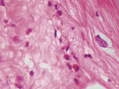
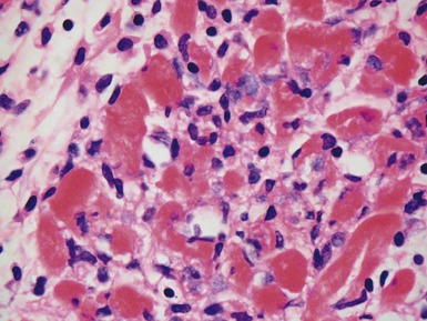
Fig 13.8 Photomicrograph of myocardium from a 12-year-old boy with a strong family history of restrictive cardiomyopathy. Many of the myocytes contain rounded brightly eosinophilic homogenous cytoplasmic inclusions that ultrastructurally are composed of aggregated filaments. In longitudinal sections the inclusions are elongated and wavy. (H&E)
VIRAL MYOCARDITIS
Histopathological features (Figs 13.9–13.13)

Fig 13.9 Photomicrograph of an 8-day-old boy with positive PCR for Enterovirus in myocardium. There is a heavy inflammatory cell infiltrate with loss of myocytes. Lymphocytes predominate but eosinophils and neutrophils are also present.

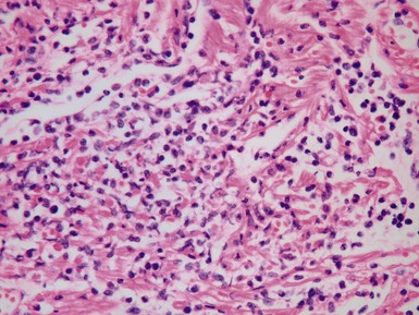
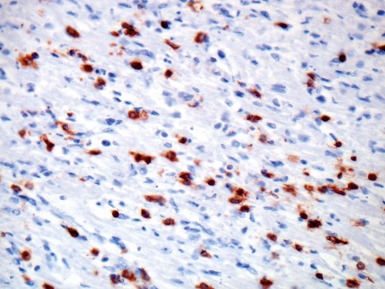
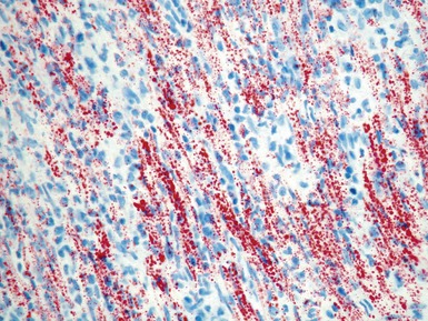
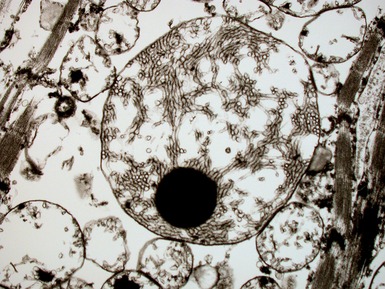
Fig 13.10 Same case as Fig 13.9 stained with anti-CD3 antibody. There are large numbers of positive T-cells in the myocardium.

Fig 13.11 Same case as Fig 13.9 stained with anti-CD79a antibody. There are only scattered B-cells.

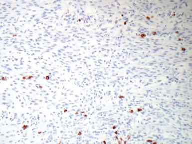
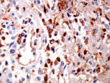
Fig 13.12 Same case as Fig 13.9 stained with antibody to CD68. There are large numbers of macrophages in the inflammatory cell infiltrate.
Differential diagnosis and pitfalls

Fig 13.14 Same case as Fig 13.9 stained for fat. Note extensive accumulation of lipid droplets in the myocyte cytoplasm. In this context, the finding of abundant cytoplasmic lipid does not indicate a disorder of fatty acid oxidation. (Frozen section stained with Oil-red-O)
NON-VIRAL MYOCARDITIS
Giant cell myocarditis (Laufs et al 2002, Das et al 2006)


Fig 13.15 Photomicrograph of heart from an 8-year-old boy with severe cardiomyopathy. Biopsy at the time of insertion of ventricular assist device. There is giant cell myocarditis. The giant cells are multinucleate and there is a heavy background lymphocytic infiltrate as well as extensive myocyte destruction. Contraction bands are evident in the lower field.
ASSESSMENT OF THE EXPLANTED HEART
INTRODUCTION
CARDIOMYOPATHIES IN THE EXPLANTED HEART

Fig 13.18 Macroscopic photograph of an explanted heart cut in a simulated 4-chamber echocardiographic view. Both atria and ventricles are clearly shown. The atrioventricular junctions and valves are well seen, as is the interventricular septum, both muscular and membranous. The posterior wall of the left atrium is not included, nor are the cavo-atrial junctions; these are retained for surgical anastomosis in the patient. The great arteries and arterial valves are not seen in this view but can be inspected and sampled nonetheless.

Fig 13.19 Macroscopic photograph of an explanted heart with dilated cardiomyopathy cut in a simulated echocardiographic parasternal long-axis view. It shows a cannula inserted into the aorta and a cannula inserted into the apex of the left ventricle. The epicardium over the right ventricular wall is thickened and hemorrhagic in keeping with the surgery to insert the cannulae of the left ventricular assist device three and a half months previously.

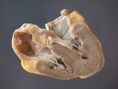
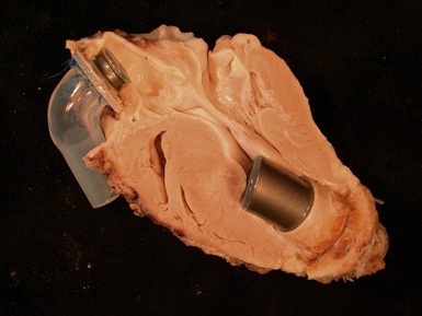
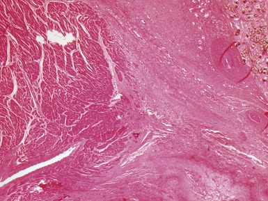
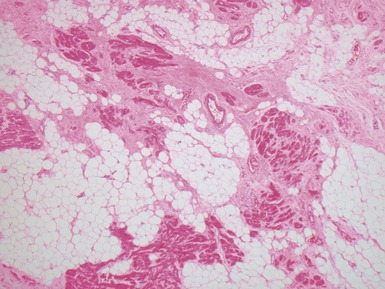
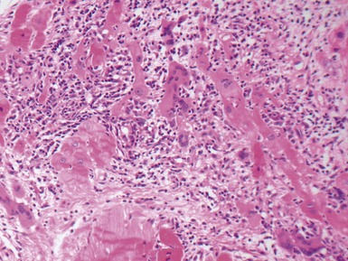
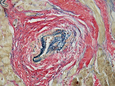
Fig 13.20 Same case as Fig 13.19, showing the cannula insertion site at the left ventricular apex. There is dense myocardial fibrosis. Dark flecks of dystrophic calcification are seen at the junction with the myocardium. Suture material is evident on the right. Inflammation is minimal in this case.
Histopathological features of cardiomyopathies in explanted hearts
Dilated cardiomyopathy
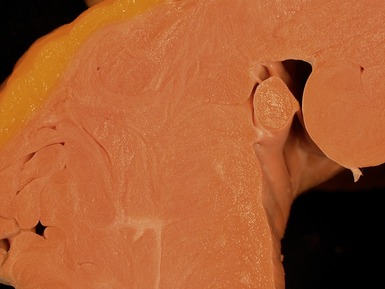
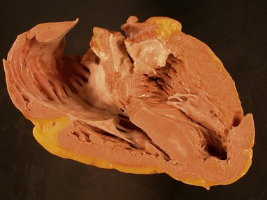
Macroscopic features (Fig 13.21)

Fig 13.21 Macroscopic photograph of a formalin-fixed, explanted heart from a 14-year-old boy with dilated cardiomyopathy. The heart is cut in a simulated echocardiographic four-chamber view. The arterial valves have been harvested before fixation. The left ventricle is dilated and globular and shows opaque endocardial thickening. There is also right ventricular and right atrial dilatation. There is stretching and thinning of the muscular trabeculae, including the papillary muscles in both ventricles.
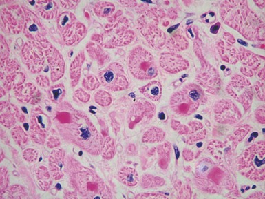
Microscopic features (Figs 13.22–13.24)

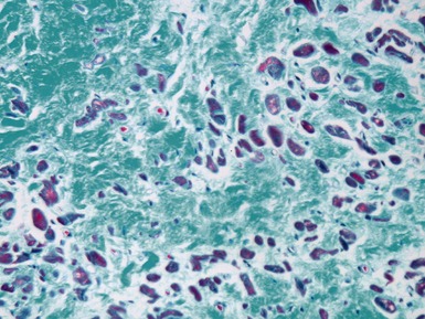
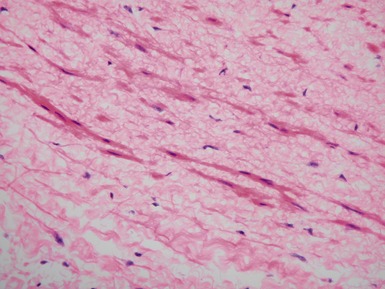
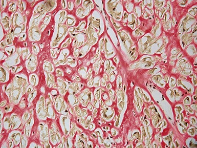
Fig 13.22 Photomicrograph of heart from a 4-year-old boy with dilated cardiomyopathy. Section of left ventricular myocardium of explanted heart shows extensive interstitial fibrosis with myocyte dropout. (Masson trichrome)

Fig 13.23 Photomicrograph of heart from a 1-year-old girl with dilated cardiomyopathy. Section of left ventricular wall shows prominent fibroelastic endocardial thickening. Note the extension of the fibroelastic tissue into the intertrabecular recesses. There is also an increase in interstitial fibrous tissue. (Elastic van Gieson)
Hypertrophic cardiomyopathy
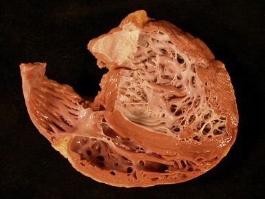
Macroscopic features (Fig 13.25)
Microscopic features (Fig 13.26)

Fig 13.26 Photomicrograph of an explanted heart from a 15-year-old boy with hypertrophic cardiomyopathy. The section from the interventricular septum shows a dysplastic coronary artery; there is increased adventitial collagen, the medial muscle coat is irregular and thin and there is irregular intimal thickening by fibroelastic tissue with luminal narrowing. The appearance is typical of hypertrophic cardiomyopathy. (Elastic van Gieson)
Restrictive cardiomyopathy
Macroscopic features (Fig 13.27)

Fig 13.27 Macroscopic photograph of the explanted heart from a 14-year-old boy with restrictive cardiomyopathy. The arterial valves were harvested before fixation. The heart is cut in a simulated echocardiographic four-chamber view. Note the thickening of the ventricular myocardium, the fibrosis in the center of the interventricular septum and the marked dilatation of the right atrium.
MITOCHONDRIAL CARDIOMYOPATHIES
Histopathological features

Fig 13.29 Same case as Fig 13.6. Electron photomicrograph of a cardiac myocyte showing a huge mitochondrion with tubular cristae and dense inclusion.
ARRHYTHMOGENIC RIGHT VENTRICULAR CARDIOMYOPATHY

Stay updated, free articles. Join our Telegram channel

Full access? Get Clinical Tree

