chapter 10 Cardiovascular Assessment of Infants and Children
Whatever you do, do it to the purpose; do it thoroughly, not superficially. Go to the bottom of things. Anything half done, or half known, is in my mind, neither done nor known at all. Nay, worse, for it often misleads.
Cardiology, to a degree perhaps seen in no other subspecialty, relies heavily on the clinical skills of practitioners to identify disease. This reliance on clinical skills has not changed, even in this age of advancing and readily available technology. Cardiovascular assessment of children requires patience, thoroughness, and flexibility, because often you must adapt to children who are in varying states of arousal and may not be at all ready for you to examine them. It also requires a great deal of compassion, because few things engender as much parental anxiety as the thought that something may be wrong with their child’s heart.
In this chapter, we will review an approach to the history and physical examination of children at different ages. Because disease states may surface at different times during childhood, and because the cardiovascular system changes with age, clinical symptoms to be explored and the approach to the physical examination are described separately for four age groups: the infant, the 3- to 5-year-old child, the school-aged child, and the teenager. A systematic approach to history-taking and physical examination helps you develop the skills and confidence that will allow you to make correct decisions for most children without resorting to indiscriminate use of imaging or other investigations.
Classification of Heart Disease in Children
When taking a history of a presenting complaint with a potential cardiac cause or association, remember that heart disease in children takes several forms. These types of heart disease may be divided into the following categories:
Thus, in approaching the cardiovascular examination of the child, be aware not only of the variations of normal but also of the wide spectrum of diseases that may occur.
Peter is a 3-month-old who is brought to the office by his mother because of new concerns about feeding. In the past 2 weeks, his feeding has decreased significantly. He is irritable and upset a lot of the time and seems to be constantly clammy or sweaty. His mother thinks he also breathes faster than other babies. She has never noticed him to look looks blue, but her friends comment on how pale he is. Her husband, who works on an oil rig offshore, thinks the baby has colic and that she is overreacting.
The experienced clinician will recognize these symptoms as potentially worrisome and highly suggestive of congestive heart failure. Further history-taking that explores the details around his symptoms, any associated symptoms, and the perinatal and family history will help narrow down potential causes. A careful, thorough physical examination will help confirm suspicions and allow appropriate investigation, referral, and timely treatment.
In this case, the timing of the onset of symptoms is slightly later than is usually seen in lesions, such as VSDs or atrioventricular (AV) septal defects (AV canals), which are the most common causes of congestive heart failure in infancy. These lesions usually become symptomatic between 4 and 6 weeks of life, as pulmonary vascular resistance falls enough to allow enough left-to-right shunting to cause congestive heart failure. Given the later onset and the irritability in this child, a more unusual lesion also must be considered—an aberrant left coronary off the pulmonary artery. These lesions “steal” blood away from the left coronary system and result in both a dilated cardiomyopathy and recurrent angina for the baby. Correction of this problem can be accomplished surgically but is much more urgent than correction of a ventricular level shunt.
The assessment of infants and children who present with symptoms suggestive of heart disease is described in this chapter. Tips for separating “functional” or benign symptoms or physical examination findings from more serious ones are also provided.
History of Present Illness and Cardiac Functional Inquiry in the Infant and Young Child
Of every 1000 live births, approximately 13 infants are born with a congenital cardiovascular anomaly. Hence, when evaluating newborns and infants for potential cardiac problems, congenital heart disease should be at the top of your differential diagnoses. Less common conditions that may arise are persistent pulmonary hypertension, asphyxia, and symptomatic cardiac arrhythmias.
Congenital heart lesions can be divided into three groups:
The three most common clinical presentations of heart disease in a newborn or infant are (1) a murmur, (2) cyanosis, and (3) respiratory difficulty.
To help identify the underlying pathology, always interpret clinical findings in terms of the underlying hemodynamic disturbance, as described in the following discussions of clinical manifestations.
Respiratory distress
When a newborn or a young infant is in respiratory distress, do not assume that the underlying problem is primarily respiratory. A child whose problem is primarily cardiac may present with pulmonary symptoms or secondary pulmonary complications, such as an infection.
For our purposes, two types of respiratory distress can be defined: (1) tachypnea, or abnormally rapid respirations, and (2) dyspnea, or difficulty breathing.
Cyanotic heart lesions or lesions involving low cardiac output may be associated with a compensatory rapid respiratory rate, particularly on exertion, because of diminished peripheral oxygenation. Children with cyanotic lesions often are tachypneic without displaying increased work of breathing (i.e., dyspnea).
Left ventricular (LV) failure from any cause results in a high end-diastolic pressure in the left ventricle and elevated pulmonary venous pressure. This in turn causes a higher back pressure in the pulmonary vessels and transudation of fluid into the pulmonary interstitial spaces, making the lungs less compliant. The child then works harder to breathe and becomes dyspneic. To assist with the increased work required for breathing, the accessory muscles come into use, and subcostal indrawing is observed. Respirations become rapid. The child’s wet, stiff lungs are very susceptible to secondary infection, which may be the reason medical attention initially is sought for the patient.
Fatigue, excessive perspiration, and poor weight gain
In young infants, metabolic demands are usually greatest during feedings. Thus the infant with poor peripheral oxygenation as a result of low cardiac output tires easily during feeding (the equivalent of exercise in older children). Because of fatigue, the infant is unable to take a full feeding. In addition, rapid respiration diminishes the time available for swallowing. This combination of factors results in failure to gain weight. In the baby with a large left-to-right shunt, such as a VSD, the process is exaggerated by the higher caloric needs of an overworked myocardium. Increased sympathetic activity causes excessive perspiration, which often is a valuable diagnostic feature.
It also is important to ask about the duration and quantity of feedings. Feeding usually is accomplished within 20 minutes, which means that the mother’s breast usually should feel empty by then or the bottle should be finished. In a bottle-fed baby, the amount of formula taken per feed and how many times per day the baby feeds are important details that may help you understand the baby’s difficulties and can be used in guiding future treatment.
Squatting
Parents of older children with certain cyanotic heart defects, especially tetralogy of Fallot, may offer the observation that when their youngster tires, he or she assumes a squatting position. Squatting helps increase systemic oxygen saturation by decreasing the amount of right-to-left shunting. These days squatting is a very uncommon symptom/sign in the developed world because most children with conditions for which squatting helps have their lesions fully repaired long before they are able to walk.
Cyanosis
Parents frequently report that their children turn blue. On further inquiry, this phenomenon turns out to be blueness of the hands and feet only, or blueness around the lips. If the lips themselves remain pink, all of these events represent peripheral cyanosis or acrocyanosis. In some disease states, this phenomenon can represent a condition in which there is decreased peripheral circulation as a result of poor ventricular function, although children with that type of problem generally look unwell in other ways. Much more commonly, acrocyanosis represents immaturity of the autonomic nervous system, causing peripheral vasoconstriction and slower peripheral circulation as a result. Consequently, acrocyanosis in a child who is well should be considered benign.
In contrast, central cyanosis occurs when more than 5 g/dL of deoxygenated hemoglobin is present in the blood. This condition is most commonly manifested as blueness or sometimes as excessive redness or as a purple tinge of the lips and tongue. Although central cyanosis can come from a variety of cardiac, respiratory, neurologic (hypoventilation), or hematologic causes, its presence is always abnormal.
Hypercyanotic spells
In some forms of congenital heart disease, the degree of cyanosis worsens periodically and can be quite profound, to the point of causing loss of consciousness. These episodes are known as hypercyanotic spells and occur classically in children with cyanotic congenital heart disease that involves narrowing of the infundibulum (the subpulmonary outflow tract) and a VSD, classically tetralogy of Fallot. This clinical phenomenon is caused by either: (1) an increase in pulmonary resistance (resulting from either a subpulmonary infundibular muscle spasm or an increase in peripheral pulmonary vascular resistance) or (2) a fall in systemic vascular resistance. Both mechanisms result in a relaive increase in the restriction of blood flow out of the right ventricular outflow and a subsequent increase in right-to-left shunting across the VSD. Spells often are precipitated by crying. After patients with hypercyanotic spells have been stabilized, their condition should always be discussed with a pediatric cardiologist before they are discharged from the hospital or emergency department.
Angina
Angina is rare but not unknown in infants and children; it can occur in persons with severe aortic stenosis or possibly pulmonary stenosis because of associated myocardial ischemia. It also may occur in persons with certain forms of congenital heart disease after surgical repair that involved manipulation of the coronary arteries. Angina may be seen in patients with Kawasaki disease in whom coronary stenoses or thromboses develop within coronary aneurysms. Angina has been recognized in infants with very rapid paroxysmal tachycardias and in those with an aberrant left coronary artery. Rarely, children with homozygous forms of familial hypercholesterolemia can have angina as a result of atherosclerotic coronary narrowing. Chest pain in this rare group of patients must always be taken seriously.
The manifestations of angina in young children are varied but may include classic chest pain or simply periodic irritability associated with sweating or pallor. When chest pain in children is invariably associated with exercise, angina should be a consideration.
Peripheral edema
Infants and young children differ strikingly from adults regarding the development of peripheral edema in the presence of congestive heart failure. Pretibial and presacral edema are late developments in a child with congestive circulatory failure. This phenomenon is believed to be due to a difference in tissue turgor. When peripheral edema resulting from heart failure does develop in an infant, it first appears periorbitally and usually is preceded by other manifestations, such as tachypnea, tachycardia, dyspnea, and liver enlargement.
Orthopnea
Unlike in adults, orthopnea is not obvious in the infant with heart failure, even when tachypnea, dyspnea, hepatomegaly, and the radiographic findings of pulmonary edema are present. In the adult, orthopnea is a symptom; in the infant, it is a sign.
Age of onset
Significance of the Age of Onset of Congestive Heart Failure
The clinical significance of the age of onset of congestive heart failure is as follows:
Significance of Age of Onset of Cyanosis
Central cyanosis resulting from congenital heart disease may be present at birth or may first appear when the ductus arteriosus closes off, usually by 5 days after birth. In persons with tetralogy of Fallot, it may develop later when the infundibular stenosis becomes more severe, increasing the volume of right-to-left shunting. Because the murmur of infundibular pulmonary stenosis greatly resembles that of a small VSD, the newborn infant presenting with what appears to be VSD could just have tetralogy of Fallot.
Family history
Genetic factors are increasingly recognized to be important in the etiology of congenital heart disease. Reports exist of as many as three children of the same parents being affected with VSD, PDA, and aortic stenosis. These are exceptions, however, because the tendency for congenital heart disease to occur in a child when one parent has a congenital heart lesion is only mildly increased (2% to 4% in most cases, compared with about 1% in the general population). The genetic tendency for a congenital heart lesion also exists in siblings, particularly for left-sided lesions such as bicuspid aortic valve. In certain syndromes or hereditary disorders with cardiac implications, however, the genetic tendency for recurrence is high. For example, Marfan syndrome is inherited as an autosomal dominant gene, and thus there is a 50% chance of recurrence. The same is true in 22q.11 deletion syndromes, which are highly associated with conotruncal cardiac defects. Consanguinity is a significant causative factor in congenital heart disease, but the tendency varies between families and with the type of heart lesion.
In summary, a search for evidence of congenital heart disease in the family is important. However, when there is a history of congenital heart disease in one parent or in a previous child, counseling regarding the risk of recurrence is best done by a geneticist.
Prenatal history
Because the cause of congenital heart disease is multifactorial, known contributory factors should be sought in the prenatal history, including:
In most instances, however, no specific contributory factors can be identified.
History of delivery
An important but infrequent cardiovascular problem in newborns is persistent pulmonary hypertension, which may cause central cyanosis, myocardial dysfunction, or both. This condition often is preceded by a difficult delivery and meconium aspiration. It is unlikely to occur after an uncomplicated delivery. Clinical differentiation of pulmonary hypertension from congenital heart disease may be difficult and usually requires echocardiography.
Determining the gestational age of a newborn also is very important because persistent patency of the ductus arteriosus is common in premature infants.
Approach to Cardiovascular Examination of the Infant
Because infants and children have an unfortunate habit of not always cooperating, you need to have an organized approach to the cardiovascular examination, while also staying flexible. Do what can be done when the opportunity arises. Begin by assessing the child’s physical development and looking for dysmorphic features, using a systematic approach (see Chapter 5).
Infants and toddlers usually are most comfortable on the parent’s lap during the examination. Taking a minute to play a game such as peek-a-boo at the start of the examination of a toddler often goes a long way toward winning the trust of the child, and it pays dividends in the cooperation you get later during the examination.
Initial assessment
When you start the examination, don’t undress the baby right away. Begin with something relatively non-threatening, such as an examination of the hands. The baby usually allows you to examine the palmar creases and to check the nail beds and muscle tone without much protest. Then feel the brachial pulses for rate, rhythm, and volume, the last being the most important. Feel this pulse in every baby you examine to learn the difference between a normal and abnormal brachial pulse. An abnormally “full” pulse suggests a PDA or aortic insufficiency. In premature infants, palpable palmar pulses (from the palmar arch) indicate the same thing. A shallow, slow-rising brachial pulse suggests LV outflow tract obstruction. Do not feel for the femoral pulses yet.
Looking for central cyanosis
Because the most pressing clinical problems are congestive heart failure and cyanosis, decide early in the examination whether central cyanosis is present. Because this determination is not always easy, you may find an experienced nurse’s opinion invaluable. Many normal newborns have a deep plethoric appearance as a result of a transiently high hemoglobin concentration, particularly if there was a delay in clamping the umbilical cord. Plethora is not as obvious in the mucous membranes, so look carefully in the baby’s mouth. Deep pressure on the skin may help, because the blanched area does not pink up as quickly in infants with central cyanosis. Many normal infants exhibit a generalized mottling, particularly after being bathed (see Fig. 4-2); this condition is called cutis marmorata (literally, marbled skin) and is not pathologic. It also is common in children with Down syndrome. Observe the effect of crying. Invariably, central cyanosis resulting from cardiac disease increases during crying, but do not make the baby cry until after you have listened to the heart.
Clinical manifestations of heart failure
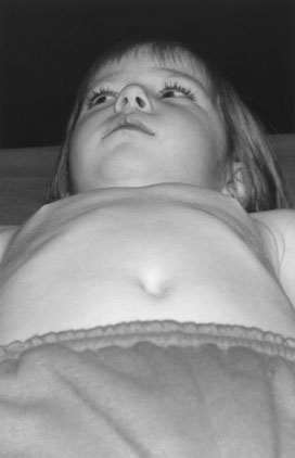
When low cardiac output and high pulmonary venous pressure cause sufficient hemodynamic disturbance to produce clinical manifestations, cardiac enlargement is invariably present. Whether the disturbance involves primarily the left or the right ventricle, the left side of the thorax becomes prominent anteriorly (Fig. 10–1). Although this prominence may not be evident in the first month of life, it certainly will be evident by the age of 3 months. When respiratory distress due to heart failure has been present for 2 months or longer, the greater diaphragmatic contractions during respiration may produce a sulcus in the lower thorax, with outward flaring of the inferior rib cage edge. Therefore, look for a sulcus, left-sided chest prominence, abnormal chest or abdominal movement, an increased respiratory rate, and subcostal indrawing.

FIGURE 10–1 Prominence of the left side of the chest in a 3-year-old with ventricular septal defect and moderate left-to-right shunt that is causing enlargement of both left and right ventricles.
Remember that young infants are normally abdominal breathers, so you must be certain that you are not simply observing normal chest-abdominal movement. Also, be sure that the indrawing is not restricted to the midline, as occurs in persons with pectus excavatum. True subcostal indrawing is abnormal and usually signifies stiffness of the lungs from either cardiac or pulmonary causes. In contrast to adults, in young children examination of the jugular venous pulse for evidence of volume overload is useless.
Palpation
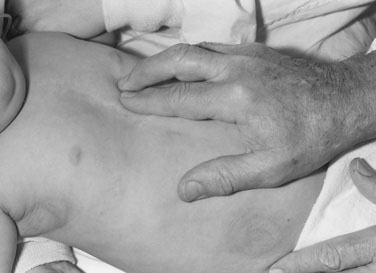
Now lay your prewarmed hand very gently on the infant’s chest, remembering that the heart may not be in its normal position. With the tips of the first and second fingers of your right hand, depress the thorax just left of the xiphoid process (Fig. 10–2). Your fingertips are now lying on the right ventricle. A faint impulse is allowable, but if the heart is enlarged, a definite forceful movement will be present. When you perform this maneuver repeatedly in normal infants, you soon will be able to tell the difference between normal and abnormal findings. This distinction will help you make a quick decision about whether the 6-week-old baby who presents with respiratory distress has a cardiac or a respiratory problem.

FIGURE 10–2 Press on the precordium to the left of the xiphoid process with the first and second digits of your right hand to detect enlargement of the right ventricle.
Except in the rare instance in which the baby has a dilated cardiomyopathy, if the respiratory distress is due to heart failure, a prominent precordial pulsation is evident. It is that simple.
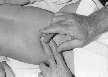
Now depress the thorax in the apical area. Prominence of the apical impulse is diagnostically less helpful in infants than in older children, except in rare instances, such as in tricuspid atresia in which the right ventricle is hypoplastic. Then palpate in the second interspace at the left sternal border, where a prominent pulmonary artery pulsation may be elicited. Finally, place one index finger carefully in the suprasternal notch (Fig. 10–3), searching first for an abnormal pulsation and then for a thrill. Then work in the opposite direction, searching for thrills and palpable sounds. At this point, you should have made a reasonable appraisal of the child’s cardiac dynamics.
Liver size and position
Whether you are right- or left-handed, stand or sit on the baby’s right side to palpate for the liver. Use the tip of your right thumb and begin well down in the right lower quadrant of the abdomen, pressing inward and upward (Fig. 10–4). If the baby has just been fed, do not press very deeply. If the edge of the liver is soft, its margin may be difficult to detect; nevertheless, if the liver is enlarged, you should appreciate a sense of resistance as your thumb tip moves superiorly. If the edge is difficult to feel, use soft percussion, tapping the second digit of your left hand with the second digit of your right hand, beginning low in the right lower quadrant and placing the second digit of your left hand parallel to the liver edge (Fig. 10–5). You should be able to sense the change in the percussion note signifying the liver’s edge. Except in the presence of pulmonary hyperinflation, the edge of the liver normally should not be more than 1 to 2 cm below the costal margin.

FIGURE 10–4 Palpate the liver with movement of the tip of the thumb inward and cephalad, beginning low in the right lower quadrant of the abdomen.

FIGURE 10–5 Percuss for the liver edge, using soft percussion with the second digit of your right hand on the second digit of your left hand, which has been positioned parallel to the liver edge.
In the presence of heart failure, the liver will be enlarged; therefore, if you find that the heart action is increased and the liver is enlarged upon palpation, you can be sure that the baby has a serious cardiac problem, even before you have used the stethoscope.
Finally, remember that the liver can be ectopic (i.e., on the left side or up in the thorax).
Auscultation
You need all the acoustic help you can get during cardiac auscultation, so be sure to turn off radios and televisions, close the door, and have everything and everyone be as quiet as possible. Cardiac auscultation is not easy, even in older cooperative patients, but coping with a restless baby with rapid cardiac and respiratory rates in a noisy nursery is a real trial. Having the child eat or giving him or her a pacifier may help. Hospital stethoscopes frequently are of poor quality, so find and use a good one.
Remember that the two main determinants of auscultatory proficiency are the fit of the earpieces and the quality (or at least the education!) of the gray matter between them.
Recognition of normal splitting of the second heart sound often is impossible when the heart rate is rapid. However, you should be able to assess the intensity or loudness of the second sound. Its intensity increases in the presence of pulmonary hypertension or when the aorta is anteriorly placed, as in transposition of the great vessels. Occasionally an ejection sound can be appreciated, which is an abnormal finding. Listen over the back for the murmur of coarctation and to both sides of the skull for the bruit of an intracranial arteriovenous malformation.
Breath sounds often interfere with the interpretation of heart sounds; however, most babies will stop breathing for a couple of seconds after you administer a surreptitious puff of air in the face or briefly and gently pinch the nose.
A detailed description of cardiac auscultation and the types of findings possible is contained in a subsequent section of this chapter. The following list contains a few dogmatic but valuable generalizations concerning auscultatory findings in young infants:
Palpating the femoral pulses
Palpation of the pulses calls for gentleness, persistence, and patience, so make yourself comfortable before you begin. First, remove the baby’s diaper and palpate for femoral pulsations. Many babies do not appreciate having people poke around in their groins and may cry, urinate, or both. Femoral pulses are particularly difficult to appreciate in obese babies; thus, do not rush into a diagnosis of coarctation of the aorta if you have difficulty feeling them. If the femoral pulses are not palpable in an asthenic baby, however, there is cause for concern (see Fig. 4-20). Now palpate both brachial pulses again. If you can detect good brachial pulses and are certain that the femoral pulses are absent or greatly depressed, listen to the heart before measuring the blood pressure and upsetting the baby. Listen particularly for a high-pitched blowing systolic murmur, which is best heard anteriorly below the left clavicle and can be heard clearly in the left axilla and back, medial to the scapula.
Blood pressure
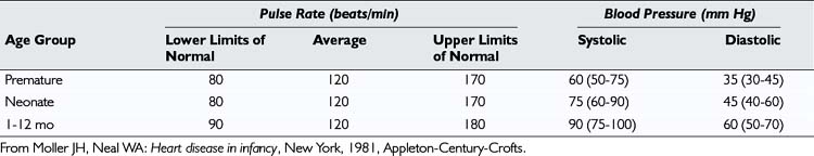
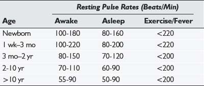
Although measuring the systemic blood pressure can be a difficult task, you should try to do so. The normal systolic blood pressure of an infant is between 60 and 80 mm Hg in both the arm and the leg (Table 10–1). The four methods of measuring blood pressure are (1) auscultatory, (2) palpatory, (3) visual (flush), and (4) Doppler.
All methods require the use of an inflatable cuff. The first decision is to choose the size of the cuff; the size is of great importance because the measuring the pressure involves occlusion of the arterial pulse (or the brachial arterial pulse when the arm is used). If the cuff used is too small, greater pressure must be used to obliterate the pulse, and the blood pressure measured will be artificially high. Use a cuff that covers almost the full extent of the upper arm, with the elbow bent. Always have a full selection of cuff sizes available. The right arm should be chosen when possible because of the proximity of the LSCA to a coarctation.
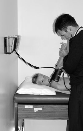
For all methods of measuring blood pressure, first supinate the child’s arm to make the radial artery easily accessible. Apply the cuff, elevate the arm, and then inflate the cuff. Prior elevation of the arm (or opening and closing the hand) prevents the auscultatory gap phenomenon (Fig. 10–6). If this procedure is not followed, when you inflate the cuff and listen for Korotkoff sounds, as you decrease the pressure in the cuff you may hear a sound appear, then disappear, and then reappear as the pressure is further decreased. This phenomenon, known as the auscultatory gap, occurs because of increased vascular resistance distal to the cuff.

FIGURE 10–6 Supinate the patient’s hand and elevate the arm before expanding the sphygmomanometer cuff. This maneuver positions the radial artery properly and eliminates the auscultatory gap.
The conventional method of measuring blood pressure is the auscultatory method. The edge of the diaphragm of a warm stethoscope is placed under the inferior edge of the cuff, and the cuff is inflated. Listen for the Korotkoff sounds as the cuff pressure is decreased, watching the sphygmomanometer needle sphygmomanometer as you listen. The first sound you hear denotes the systolic level. As the cuff pressure is decreased further, an abrupt change in the intensity of the sound may be heard. If this change is detected, it is recorded as the diastolic level. Continue decreasing the cuff pressure, recording the disappearance of Korotkoff sounds, which is recorded as the diastolic level if no intensity change has been detected. Usually two or three recordings are made, elevating the arm before each attempt.
It may be impossible to measure the blood pressure by the auscultatory method, particularly when a baby does not cooperate. The palpatory method of measurement should then be attempted. Prior elevation of the arm is not required. The radial artery pulse is palpated, and the cuff is elevated until the pulse disappears. The cuff pressure is then decreased, and the level of systolic pressure is estimated by the time of the reappearance of the pulse. Only the systolic pressure can be measured by the palpatory method. A variation of this method involves detection of the appearance of the pulse with a manual Doppler probe held on the patient’s radial or brachial pulse. This method is the one we prefer using for infants, in whom cooperation for auscultation is so frequently limited.
The flush method also measures systolic pressure only, and unfortunately, it requires two persons. Using both hands on the child’s upraised arm, express most of the blood from the arm. This maneuver sometimes can be accomplished by wrapping the arm tightly from the fingers to the cuff level with a tensor bandage. The second person should then inflate the cuff and unwrap the bandage. The arm should look relatively pale. One person watches the arm, and the other watches the sphygmomanometer. As the cuff pressure is decreased, the person watching the arm indicates verbally the moment at which the flush occurs while the other person records the manometer level.
Blood pressure measurement with automatic Doppler equipment is much easier than with the other methods, because no auscultation is required. Unfortunately, the equipment is not always available, blood pressures obtained are sometimes questionable, and the choice of cuffs may be limited.
One way or another, a reliable blood pressure measurement must be obtained. If you suspect an aortic coarctation (i.e., if there is high pressure in the arm, an absence of femoral pulses, and a murmur), repeat the procedure in the thigh, using a blood pressure cuff of an appropriately larger size. Again, the manual Doppler method with the probe held on the posterior tibial or popliteal pulse is our preferred method of measuring blood pressure. The normal range of systolic and diastolic blood pressures for older children is shown in Table 10–2.

Stay updated, free articles. Join our Telegram channel

Full access? Get Clinical Tree

