CHAPTER 4 Branchial Anomalies
Step 1: Surgical Anatomy
♦ The course of branchial cysts, sinuses, and fistulas depend on the branchial arch from which they are derived (first, second, third, or fourth).
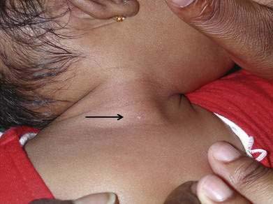
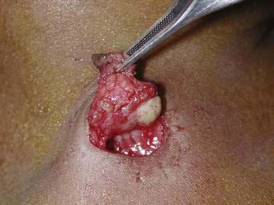
♦ The extent of branchial anomalies varies considerably, from small sinuses or cysts limited to the subcutaneous tissue (Figs. 4-1 and 4-2) to large inflammatory masses extending to the pharynx.
♦ First branchial anomalies appear in the preauricular or submandibular area and may course through the parotid gland, insinuate around the facial nerve trunk or branches, and connect to the external auditory canal.
♦ Second branchial anomalies are seen along the anterior border of the sternocleidomastoid muscle and may ascend through the deep tissues of the neck above the hyoid, between the internal and external carotid arteries, adjacent to the hypoglossal and glossopharyngeal nerves, and terminate in the tonsillar fossa or other nasopharyngeal areas.
♦ Although the theoretical course of third and fourth branchial anomalies has been described as descending into the mediastinum, this course has not been noted in vivo. These rare lesions have more recently been described by their pharyngeal terminus as pyriform fossa sinuses. Externally they may be evident along the anterior border of the sternocleidomastoid, usually on the left side of the neck, and pass through the thyroid, posterior to the internal carotid, adjacent to the recurrent laryngeal nerve, through the inferior constrictor muscle, and connecting to the pyriform fossa.
♦ Branchial anomalies may contain keratinizing or nonkeratinizing squamous epithelium or pseudostratified columnar respiratory epithelium.
Step 2: Preoperative Considerations
♦ Most common initial signs of branchial anomalies are an asymptomatic cystic mass, chronically draining sinus, or recurrently infected lesion of the neck.
♦ Occasionally it may manifest in infants with respiratory distress or stridor resulting from the swelling of cyst, impinging on the airway.
♦ Preoperative imaging may help distinguish a branchial anomaly from other lesions, such as a cystic hygroma, lymphoma, neurofibromatosis, lymphadenopathy, carotid body tumor, tuberculous adenitis, lipoma, hemangioma, thyroglossal duct cyst, ectopic thyroid, thymic cyst, or a dermoid.
♦ Ultrasound or computed tomography scan with intravenous contrast may demonstrate gas in a cystic structure and may be useful to delineate the course of the lesion and possible thyroid involvement.
♦ Antibiotics (with drainage, if necessary) should be administered until clinical signs of infection have resolved and before removal to minimize risks of complications and recurrence.
♦ Removal of a branchial anomaly before repeat infections will aid dissection in clean surgical planes.
♦ Laryngoscopy or telescopic pharyngoscopy is useful for identification or cannulation of a pharyngeal fistula.
♦ Recent studies suggest that chemical or thermal cauterization of the internal opening may be adequate treatment for pyriform fossa sinus anomalies.
Step 3: Operative Steps
First Branchial Anomalies
♦ Injection of the tract through the external auditory canal with methylene blue or instrumentation with a small probe facilitates tract identification.
♦ The superficial lobe of the parotid is mobilized with clear identification of the facial nerve as it emerges from the stylomastoid foramen and enters the parotid.
Stay updated, free articles. Join our Telegram channel

Full access? Get Clinical Tree


