60 Bone Neoplasms
Osteosarcoma
Clinical Presentation
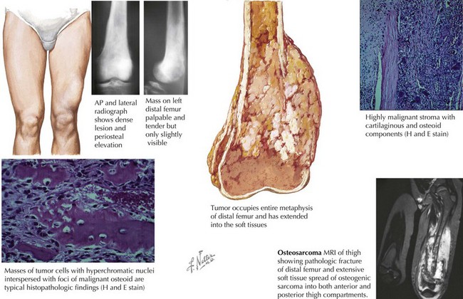
The most common clinical symptom at presentation is pain, often described as dull and aching, and typically of several months’ duration. Other complaints include a palpable mass with or without swelling. Systemic complaints such as fevers, weight loss, and decreased appetite are rare. Eighty percent of OS occur in the extremities, and on examination, a mass (tender or nontender) may be noted (Figure 60-1). The examination may also reveal decreased range of motion or muscle atrophy. Regional lymphadenopathy is rare. The most common sites of disease include the distal femur, proximal tibia, and proximal humerus, although OS may occur in any bone. Involvement of the axial skeleton can occur but is less common. Eighty percent of patients with OS have localized disease at the time of diagnosis. The lung is the most common site of detectable metastatic disease at diagnosis, although patients may present with multifocal bone disease without pulmonary involvement.
Evaluation
Imaging studies are more helpful in making the diagnosis of OS than are laboratory tests. A plain radiograph will often reveal a lytic or blastic lesion of the bone with poorly defined borders (see Figure 60-1). Other findings include periosteal elevation adjacent to the primary lesion, a sunburst appearance, or a pathologic fracture. If OS is suspected, chest computed tomography (CT) and bone scan can be used to assess for pulmonary and bone metastases. Magnetic resonance imaging (MRI) should be performed to better evaluate the extent of the tumor and should include the joint above and below the involved area so that skip lesions are not missed (see Figure 60-1). In addition to delineating the intra- and extraosseous extent of the tumor, the MRI may provide information regarding tumor effects on critical neurovascular structures.
Under the microscope, OS classically appears to be composed of spindle cells associated with malignant osteoid (see Figure 60-1). The extent of osteoid production may vary among the osteoblastic, chondroblastic, fibroblastic, telangiectatic, and small cell subtypes; however, the presence of tumor osteoid is the key pathologic feature of this disease.
< div class='tao-gold-member'>
Stay updated, free articles. Join our Telegram channel

Full access? Get Clinical Tree


