12.2 Bone and joint infections
Introduction
Infections of bones (osteomyelitis) and joints (septic arthritis) can occur at any age but are much more common in children than adults. They are often difficult to diagnose, can be difficult to treat and, if not diagnosed and treated promptly, can lead to permanent disability. Many children and adults have shortened or deformed limbs, fused joints or even amputations because osteomyelitis or septic arthritis was diagnosed too late or treated inadequately. Almost all these poor outcomes need not have happened; with careful history-taking and examination, prudent use of investigations – particularly imaging – and close observation, virtually all bone and joint infections can be diagnosed and treated before permanent damage ensues.
Pathogenesis
The area around the growth plates of children’s bones is particularly prone to infection. Although the metaphysis has a plentiful blood supply from nutrient arteries, blood flow through capillary loops and sinusoidal veins at the metaphyseal–epiphyseal junction is slow, which allows bloodborne bacteria to deposit in this region (Fig. 12.2.1). This area also has poor penetration of white blood cells and other immune mediators, so deposited bacteria are relatively protected. As the infection progresses, pus accumulates under pressure, further limiting the blood supply to the region. Increased stasis and activity of cytokines encourages clots to form in blood vessels, leading to ischaemic bone necrosis. Infection then spreads to the cortex through the Volkmann canals and haversian system, and subsequently into the subperiosteal space.

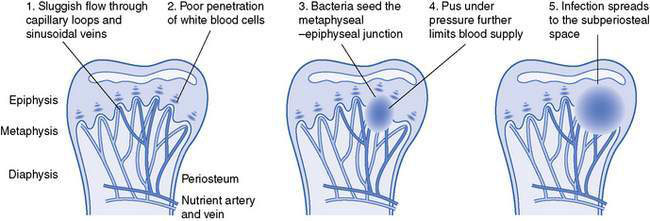
Fig. 12.2.1 Anatomical evolution of osteomyelitis.
(Adapted with permission from Krogstad P, Smith AL 2004 Osteomyelitis and septic arthritis. In: Feigin RD, Cherry JD, Demmler GJ, Kaplan SL (eds) Textbook of pediatric infectious diseases, 5th edn. WB Saunders, Philadelphia, PA, pp 683–703.)
If the infection remains untreated, bone necrosis may lead to development of a sequestrum – an area of dead cortical bone separated from normal bone. Sometimes the infection becomes walled off by granulation tissue that forms a fibrous capsule, the so-called Brodie abscess, usually located in the metaphysis and presenting subacutely with pain and tenderness, but rarely fever.
Chronic osteomyelitis is usually the result of untreated or inadequately treated acute osteomyelitis. Sequestra and Brodie abscesses are sometimes found in chronic osteomyelitis. Most cases of chronic osteomyelitis, like acute osteomyelitis, are caused by Staphylococcus aureus, but chronic presentations increase the likelihood of unusual organisms, including Mycobacterium tuberculosis, fungi and Kingella kingae.
Septic arthritis may occur de novo, as a result of deposition of bacteria in the joint. Alternatively there may be extension from adjacent osteomyelitis, which is more common in children than adults, possibly because of transport of bacteria through blood vessels that cross the epiphyseal plate. In some joints, the metaphysis is intra-articular, which means that osteomyelitis can transform directly into septic arthritis. These joints are:
Synovial joints are poor at clearing infection and the connective tissue may be damaged by enzymes released by bacteria. Initially, the inflammation results in a joint effusion, which may be purulent; if left untreated, the articular and growth cartilage can be destroyed. Longer-term complications may include dislocation, avascular necrosis of intra-articular epiphyses and joint destruction.
Microbiology
Although occasionally caused by fungi, and rarely by parasites or viruses, osteomyelitis and septic arthritis are predominantly bacterial infections, most often caused by S. aureus. The major bacterial pathogens are:
• Staphylococcus aureus (80–90% of culture positive cases) – usually methicillin-susceptible but community-acquired methicillin-resistant S. aureus (CA-MRSA) is on the rise in many places.
• Kingella kingae – recent studies using polymerase chain reaction (PCR) diagnosis suggest that this organism is a common cause of osteomyelitis and septic arthritis in young children (usually under 2 years), but is often not identified using standard laboratory cultures.
• Streptococcus pyogenes (group A streptococcus) – sometimes associated with varicella infection.
• Streptococcus pneumoniae – mainly in children aged less than 2 years.
• Pseudomonas aeruginosa – immunocompromised patients or traumatic (classical cause is a nail through a tennis shoe causing calcaneal osteomyelitis).
• Group B streptococcus or Escherichia coli – especially in neonates.
• Haemophilus influenzae type b – in unimmunized populations (mainly in developing countries).
• Mycobacterium tuberculosis – mainly in children from developing countries or immigrant populations; 50% of cases affect the spine. Often subacute presentation.
• Salmonella spp – particularly in people with sickle cell anaemia.
• Neisseria gonorrhoeae – mainly in developing countries; may cause multifocal septic arthritis in neonates or sexually active adolescents.
Clinical presentation
Although osteomyelitis and septic arthritis are usually considered distinct entities, in paediatric practice they are sometimes difficult to distinguish and may occur together. Children with septic arthritis are more likely than adults to have osteomyelitis in the adjacent bone. In neonates and infants, osteomyelitis and septic arthritis coexist so commonly that the preferred term is ‘osteoarticular infection’.
The typical child with osteomyelitis or septic arthritis is aged less than 5 years: approximately 50% of cases occur under the age of 5 years and of these 25% are under the age of 1 year. Usually the symptoms have been present for 2–3 days prior to presentation. Early in the illness, when bacteraemia is present, the child may be unwell with fever and malaise. Later, as the infection establishes itself, the symptoms relate to the main site of infection. Both diseases usually present with fever and limb pain; in younger children this will most often manifest as a limp, or disuse of a limb or other body part. Septic arthritis is more likely than osteomyelitis to have obvious localized clinical signs.
Osteomyelitis
In osteomyelitis, there is pain, which may be poorly localized, especially in young children. Neonates may present with a generalized febrile illness, without any localizing features. The precise location of the infection can often be elicited with careful physical examination, looking particularly for metaphyseal tenderness. Erythema and swelling are signs of abscess formation, which occurs late in the illness. In one-third of cases there is a history of mild preceding trauma to the affected area so such a history should increase rather than reduce the suspicion of osteomyelitis.
Osteomyelitis most commonly affects, in order:
The pelvis and vertebrae are less commonly affected but more likely to present with advanced disease because of delayed presentation (in vertebral osteomyelitis) or delayed diagnosis (in pelvic osteomyelitis). The diagnosis of osteomyelitis in long bones may also be delayed – and hence become subacute or chronic – because of formation of a Brodie abscess or the presence of low-grade infection, sometimes due to unusual organisms. In these cases, diffuse pain or tenderness is prominent and fever may be absent.
Septic arthritis
Differentiating septic arthritis from osteomyelitis can be difficult. Children with osteomyelitis may not want to move the adjacent joint because of either pain or muscle spasm. In addition to fever and limb pain, the hallmark of septic arthritis is a warm, tender joint with a dramatically restricted range of movement and an effusion. However, these signs are not always present. Septic arthritis of the shoulder or hip is very difficult to diagnose early, because of the lack of visible joint swelling in the early stages.
The following features should raise suspicion that there is coexistent osteomyelitis and septic arthritis:
Differential diagnosis
The most important diagnosis to exclude in possible osteomyelitis is malignancy. Bone tumours can cause local bone destruction, and leukaemia may present with fever and bone pain. Cellulitis may mimic the focal tenderness and erythema of late-presenting osteomyelitis. Patients with sickle cell disease may develop bone infarction, which can be difficult to differentiate from osteomyelitis.
Chronic recurrent multifocal osteomyelitis (CRMO) is a rare, non-infectious, inflammatory syndrome of unknown pathogenesis that affects children and young adults and is most common in girls. Affected children have prolonged symptoms of pain and swelling that relapse and recur. The clavicle is a classical site of involvement. Treatment with antibiotics does not alter the course of the disease but steroids and anti-inflammatory medication may provide symptomatic relief. CRMO usually resolves, although it may relapse and recur over a prolonged period (up to 15 years), and there is a danger of premature epiphyseal closure.
Septic arthritis of the hip can present similarly to transient synovitis (‘irritable hip’), which usually occurs following minor injury or a viral illness. Children with transient synovitis are usually not unwell and their joint signs are not as severe as those in children with septic arthritis, but in the early stages of either illness the diagnoses can be confused. Sometimes the only way to be sure of the diagnosis is to aspirate the joint and observe the child. It is important not to treat empirically with antibiotics without first obtaining a diagnostic specimen.
Acute joint swelling may be caused by inflammatory arthritis (e.g. juvenile chronic arthritis, inflammatory bowel disease, other connective tissue diseases), reactive arthritis (which may occur in association with a wide range of pathogens including Mycoplasma, cytomegalovirus, Epstein–Barr virus, parvovirus, hepatitis, rubella, Yersinia, Salmonella and Shigella species), rheumatic fever and Henoch–Schönlein purpura.
Rarely, osteomyelitis or septic arthritis may affect multiple bones or joints at the same time. This should raise the suspicion of a distant source of persistent bacteraemia such as endocarditis or occult abscess, and should lead to a thorough investigation for other sites of infection (e.g. heart, liver, spleen, brain, eyes). It should also raise suspicion of unusual organisms (e.g. N. gonorrhoeae) as a cause of multifocal septic arthritis or an alternative diagnosis if an organism cannot be identified (e.g. CRMO, inflammatory or reactive arthritis, or rheumatic fever).

Stay updated, free articles. Join our Telegram channel

Full access? Get Clinical Tree

