Chapter 44 Bleeding and Bruising
ETIOLOGY
What Causes Bruising and Bleeding?
A bruise is a collection of blood under the skin, usually from trauma to superficial blood vessels. Bruising and excessive bleeding without obvious trauma point to a disorder in one or more components of the hemostatic mechanism: platelets, coagulation proteins, and blood vessels.
How Does the Hemostatic Mechanism Stop Bleeding?
When blood vessels are damaged, circulating platelets are exposed to blood vessel collagen and connective tissue. Platelet interaction with these tissues and with other platelets causes release of platelet granules that stimulate adherence and aggregation of platelets. Platelets and damaged vessels also release tissue factors that initiate the conversion of prothrombin to thrombin. Once formed, thrombin interacts with fibrinogen to form fibrin monomers. Factor XIII stimulates the monomers to form fibrin polymers, which interact with the adhering platelets to form a fibrin plug that prevents further bleeding. Platelet plug formation plus contraction of the blood vessel make up the primary hemostatic mechanism.
What Platelet Problems Cause Bleeding?
Platelets may fail to function properly or may be decreased in number. Increased destruction and decreased production both result in a decrease in platelets (Table 44-1). Most disorders in both categories occur infrequently, but all must be considered carefully.
Table 44-1 Disorders Associated with Decreased Platelet Count
Increased Destruction |
Immune mediated (ITP, isoimmune) |
Disseminated intravascular coagulation (DIC) |
Sepsis |
Hemolytic uremic syndrome (HUS) or TTP |
Kasabach-Merritt syndrome (hemangiomas) |
Hypersplenism |
Decreased Production |
Viral illness |
Medications (prescription and OTC) |
Leukemia or infiltrative malignancy |
Chemotherapy drugs |
Thrombocytopenia with absent radii |
Amegakaryocytic thrombocytopenia |
Wiskott-Aldrich syndrome |
Aplastic anemia |
Congenital marrow failure syndromes |
ITP, Idiopathic thrombocytopenic purpura (immune-mediated thrombocytopenia); OTC, over the counter; TTP, thrombotic thrombocytopenic purpura.
What Causes a Decreased Platelet Count?
Immune-mediated platelet destruction, also called immune-mediated thrombocytopenia (idiopathic thrombocytopenic purpura, ITP), is the most common cause of a decreased platelet count. An antiplatelet antibody develops, usually after a viral illness, and attaches to the platelets, which are then removed from the circulation by macrophages within the spleen. In children, 80% of cases of ITP are acute, resolving within 6 months with or without treatment; 20% will become chronic, persisting longer than 6 to 12 months. Older children with ITP, especially those in whom the disorder becomes chronic, may have an autoimmune disorder.
Do Newborn Infants Develop Thrombocytopenia?
A unique variety of ITP occurs when maternally derived antiplatelet IgG crosses the placenta, leading to destruction of fetal platelets. This thrombocytopenia can occur if the mother has ITP or an autoimmune disorder, or if she lacks the PLA-1 antigen on the fetal platelets. This latter situation is termed isoimmune thrombocytopenia. The mother becomes sensitized by fetal platelets that enter into her circulation. She forms antiplatelet antibodies that cross the placenta and bind to fetal platelets, shortening their survival. Thrombocytopenia develops in the fetus, which makes vaginal delivery dangerous owing to the possibility of intracranial bleeding. The newborn’s platelet count usually begins to increase several weeks after birth as the antiplatelet antibody level falls.
What Congenital Conditions Cause Thrombocytopenia?
Thrombocytopenia caused by an isolated defect of marrow platelet production may present at birth with the syndrome of megakaryocytic thrombocytopenia and the thrombocytopenia with absent radii syndrome. When reduced platelet production from congenital disorders appears later, it is often accompanied by decreases in other blood cell lines. Fanconi’s anemia has bone marrow failure, absent radii and thumbs, short stature, and increased risk of leukemia; dyskeratosis congenita is a syndrome of bone marrow failure with cutaneous and ectodermal dysplasia; and Bloom’s syndrome includes short stature, bone marrow failure, and increased risk of leukemia. Thrombocytopenia is also associated with the Wiskott-Aldrich syndrome, an immunologic deficiency characterized by eczema, recurrent infections, T-cell function abnormalities, altered antibody response to antigens, and small platelets.
What Acquired Conditions Decrease Platelet Production?
Platelet production is reduced in aplastic anemia, leukemia, and infiltrative malignancy (e.g., neuroblastoma). Bone marrow aspirate and biopsy will identify pathology of other cell lines. Viral infections or sepsis can also suppress platelet production, as can chemotherapy and prescription and over-the-counter medicines.
What Congenital Disorders Cause Platelet Dysfunction?
von Willebrand’s disease (VWD) is an autosomal dominant disorder with variable penetrance and is the most common congenital abnormality of platelet function that is associated with increased bleeding. Bleeding occurs because the quantity or function of von Willebrand’s factor (VWF) is too low to promote the platelet-platelet and platelet-tissue interactions and the stabilization of factor VIII necessary to form a platelet plug. Another platelet disorder that causes bleeding results from congenital inability to secrete stored platelet granules, either because of a deficiency of platelet granules or because of a deficiency of arachidonic acid production. This prevents the platelet plug from forming or increasing in size.
What Causes Acquired Platelet Dysfunction?
Salicylate decreases the platelet’s ability to release granules by inhibition of the cyclooxygenase pathways and by decreased production of arachidonic acid and is the most common cause of acquired platelet dysfunction. Other drugs that inhibit platelet adherence and aggregation include ethanol, antihistamines, and dextran. Cardiopulmonary bypass also inhibits platelet function.
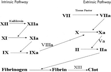
What Coagulation Problems Must I Consider?
Anything that disrupts either the intrinsic or extrinsic coagulation pathway (Figure 44-1) will result in bruising and bleeding. Congenital deficiency of factors VIII and IX are commonly referred to as hemophilia A and B, respectively. These X-linked disorders are the most common congenital abnormalities of the fluid phase of coagulation.
What Are Some Acquired Abnormalities of Coagulation?
Newborns are all deficient in the vitamin K–dependent coagulation factors because the gastrointestinal tract lacks the bacteria that synthesize vitamin K. Breastfed newborns have an additional risk because human milk has a low vitamin K concentration. To prevent vitamin K–deficiency bleeding (hemorrhagic disease of the newborn), vitamin K must be administered at birth. Bleeding is most commonly seen in newborns who were born at home and who did not receive vitamin K. Parents occasionally refuse vitamin K for hospital-born babies and must be made aware of the risk of serious bleeding. A similar bleeding problem can develop in older children if prolonged administration of antibiotics depletes the gut flora and vitamin K production falls. Sepsis can cause disseminated intravascular coagulation, which activates the hemostatic mechanism and depletes coagulation factors. Severe hepatic dysfunction can lead to decreased production of factors II, VII, IX, and X (the hepatic or vitamin K–dependent factors). Rarely, antibodies against coagulation factors develop in collagen vascular diseases and viral illnesses with resultant bleeding; paradoxically, these antibodies can also inhibit naturally occurring anticoagulants and increase the likelihood of venous thrombosis.

Stay updated, free articles. Join our Telegram channel

Full access? Get Clinical Tree

