and Paula Briggs2
(1)
Department of Obstetrics and Gynaecology, Monash University, Clayton, Victoria, Australia
(2)
Sexual and Reproductive Health, Southport and Ormskirk Hospital, Southport, UK
The basic makeup for every individual is determined by their genes. Genes are made up of a sequence of nucleotides, which are aggregated into chromosomes. The Human Genome is believed to be made up of about 23,000 genes, located in 23 pairs of chromosomes. Each species is unique in respect of the number of chromosomes that it is comprised of. The chromosomes are contained within the nucleus of every cell. In any individual the chromosomes are identical in each of its 37.2 trillion cells. As the 23,000 pairs of genes can form an infinite number of combinations and permutations (23,000 × 23,000), each individual is genetically unique with the exception of identical twins, who are formed by the splitting of an embryo, and thus start with identical genetic makeup.
Chromosome Structure
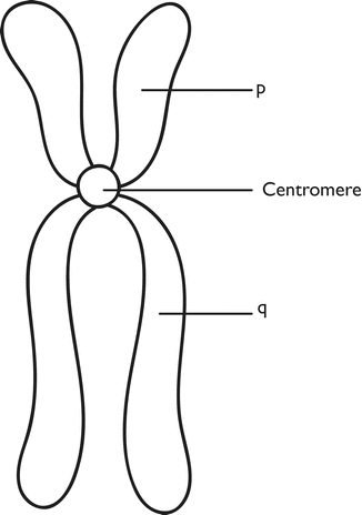
Chromosomes are a joined by a centromere. As the centromere is closer to one end, the shorter arms are called p arms and the longer arms q, and the end region is the telomere (Fig. 3.1).


Fig. 3.1
Chromosome structure
Within the nucleus, the chromosomes are in pairs, one of each pair from each parent, containing the genes present, one inherited from the male partner via the sperm and one from the female partner via the oocyte. These genes determine our characteristics. All cells of the body (somatic cells) contain these 46 (23 pairs) of chromosomes (diploid) except the germ cells (oocyte and spermatid) which contain only 23 chromosomes (haploid).
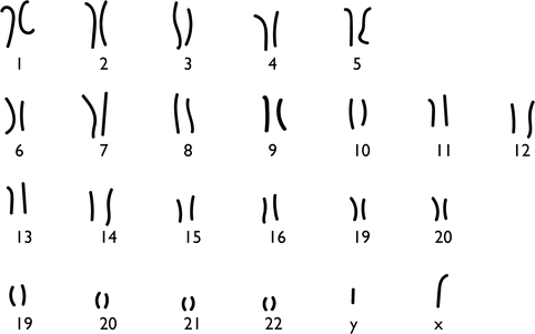
All oocytes carry 22 autosomes, with the 23rd chromosome always being an X. In contrast, sperm carry either an X or a Y bearing 23rd sex chromosome, and the 22 autosomes. Sperm that carry the Y chromosome are destined to produce a male offspring when it combines with an ovum, whereas and X bearing sperm will produce female offspring. The Y chromosome is significantly shorter than the X chromosome (Fig. 3.2).


Fig. 3.2
Karyotype
The crucial difference between germ and somatic cells is that the former reduces the diploid (46 chromosome) compliment to haploid (23 chromosome) form, thus enabling the joining of two haploid germ cells to form one new diploid individual.
During the proliferation of germ cells, we have two types of cell division, mitosis (or replication division) and meiosis (reduction division).
In the male germ cell, this reduction division occurs during its progression from the primary to secondary spermatocyte, whilst in the female from the primary oocyte to the secondary oocyte.
The female gamete is unique in that division of the primary oocyte results in uneven division of the cytoplasm, with most going to the daughter cell, the secondary ooctye, with only a very small amount going to the other daughter cell, which becomes the primary polar body. Similarly, when the secondary oocyte divides by mitosis, replicating its 23 chromosomes, one of the daughter cells again is disadvantaged with respect to the amount of cytoplasm, and it becomes the second polar body, whereas the other part becomes the mature oocyte. The polar bodies are not capable of being fertilized.
The sex of an embryo is determined at the time of fertilization and depends on whether an X bearing or Y bearing sperm has entered the ovum.
Genetic Abnormalities
Genetic abnormalities may result from loss or extra chromosomal material or an abnormality of a single gene. Chromosome abnormality may involve part of or an entire chromosome. For example Down Syndrome, where the child inherits an extra chromosome 21, known as trisomy 21. Genes can be altered in a variety of ways. The outcome depends on where the change is in the gene, how it effects gene function and how the gene change is inherited.
Single Gene Defects
Single gene defects can be dominant or recessive. With a recessive disorder, both genes (one from each parent) have to be altered in order for the abnormality to be manifest (phenotype). If only one parent has passed on the defective gene, and the same gene from the other parent is normal, the child will not be affected, but will be a carrier. In contrast, with a dominant single gene defect it is sufficient for just one gene change to be inherited, and that gene will be responsible for the phenotype, e.g. Huntington’s disease.
Stay updated, free articles. Join our Telegram channel

Full access? Get Clinical Tree


