19 Atopic Dermatitis
Atopic dermatitis (AD) is a chronic inflammatory skin disease affecting 10% to 20% of children and accounting for approximately 1 million outpatient visits per year. More than half of patients affected present before 12 months of age with an itchy rash on the face and extremities. AD is often associated with asthma, allergic rhinitis, and food allergy. The natural history of atopic disease, progressing from AD to asthma and allergic rhinitis, is often referred to as the atopic march.
Etiology and Pathogenesis
The chronic skin lesions of AD result from a complex interplay of genetic, immune, infectious, and environmental factors. In combination, these factors produce areas of persistently pruritic, inflamed skin that significantly impact the quality of life of patients. Immunologically, the systems affected most are the epidermal barrier, the first line of defense for the immune system, and the humoral immune system, which modulates the immune response to antigenic challenges by regulating antibody production.
The epidermis provides a critical barrier to keep infectious organisms, irritants, and allergens from entering the body. Genes encoding epithelial structural proteins have been strongly implicated in the pathogenesis of AD. Filaggrin is a key protein involved in keratinization that is encoded by a gene within the epidermal differentiation complex on chromosome 1q21. Defects in the gene encoding Filaggrin have been named as major causes of AD. Spink 5 is a protease inhibitor involved in intercellular attachment, which is also important in maintaining an intact epidermal barrier. It is deficient in Netherton’s syndrome, a disorder that includes severe AD.
In addition to the genes encoding epidermal barrier components, genes encoding cytokines produced by type 1 and type 2 helper T cells have also been implicated in AD. These cytokines, including interleukins (ILs) 4, 5, 12, and 13 and granulocyte-macrophage colony-stimulating factor, are important in the regulation of immunoglobulin E (IgE) synthesis. IL-18, also implicated in AD, is involved in the switching between type 1 and type 2 helper T cell responses. Children with AD have an imbalance toward a type 2 helper T cell response, resulting in a switch in B cell antibody production toward an IgE response, contributing to the development of atopic disease.
A number of other factors are important in the pathogenesis of AD. Reduced levels of ceramide, lipid molecules that make up an important component of the epidermal barrier, contribute to the compromised epidermal barrier function. The barrier is further compromised by increased proteolytic enzyme activity via increased production and a decrease in endogenous protease inhibitors. External factors, including mechanical injury from scratching affected skin, proteases from bacteria and dust mites, and the use of pH-altering soaps also contribute to a weakened skin barrier, facilitating bacterial colonization and penetration of allergens. The combination of defects in the immune system, compromised skin barrier integrity, and external pathogens and irritants sets up the cycle of chronic inflammation characteristic of AD.
Clinical Presentation
Most children with AD initially present at a very young age, with more than half presenting in the first year of life and more than two-thirds presenting before age 5 years.
History
The chief complaint of a “red, itchy rash” is common for children with AD. The child may have a pattern of similar episodes of skin eruption in a similar distribution in the past. Certain foods, clothing, soaps, lotions, or allergenic exposures (pollen, pets) exacerbate the rash of AD. There may be a history of other atopic diseases such as food allergy, asthma, or allergic rhinitis in the child or in the immediate family.
Physical Examination
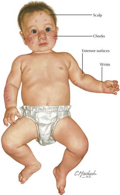
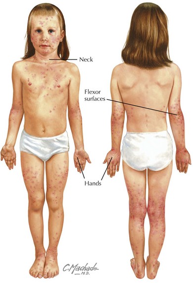
Physical examination of acute AD lesions reveals red papules and plaques that may also feature scaling, oozing, and crusting. In addition to acute lesions, children and adolescents with chronic AD may also have areas of thickened, dark skin (lichenification) at sites of prior acute lesions. The distribution of the rash is characteristic and varies by age. In infants, the cheeks, scalp, wrists, and extensor surfaces of the arms and legs are most affected (Figure 19-1). The diaper area is typically spared. The perioral and perinasal areas are rarely involved, leading to the characteristic “headlight sign” of pallor in these areas. In children and adolescents, the neck, hands, and flexor surfaces of the upper and lower extremities are more commonly affected (Figure 19-2).
Complications
The defects in the epithelial barrier leave the skin susceptible to invasion by viruses and bacteria. Two common secondary infections include superinfection with Staphylococcus aureus and eczema herpeticum.
Staphylococcus aureus Superinfection
The skin of most individuals with AD is colonized with S. aureus. Regions of the skin affected by AD are susceptible to bacterial invasion of the skin and subcutaneous soft tissues, resulting in superinfection. A local inflammatory response results with subsequent pain, erythema, edema, and warmth at the site. Serous drainage, crusting, and lymphadenopathy may be present. Fever and an elevated white blood cell count may occur if the lesions are extensive or if the bacteria have invaded the bloodstream. Surface cultures with antibiotic sensitivities should be considered to help guide appropriate therapy.
Eczema Herpeticum
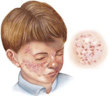
Secondary infection with herpes simplex virus (HSV) is a potentially life-threatening complication that may affect children with AD of all ages. Vesicular lesions typically appear in groups superimposed on areas of skin affected by AD between 2 days and 2 weeks after exposure to HSV-1 or HSV-2. The lesions evolve to appear as “punched-out” hemorrhagic lesions with crusting (Figure 19-3). Associated symptoms may include fever, fatigue, and lymphadenopathy. Diagnosis may be made by Tzanck prep, direct fluorescent antibody, polymerase chain reaction, or viral culture from swabs of the affected area.
Evaluation and Management
Laboratory Testing
There are no laboratory studies essential in the diagnosis of AD. If checked for other reasons, a white blood cell count with differential may reveal an eosinophilia; a serum IgE may be elevated. In severe or refractory cases of AD, an evaluation by a pediatric allergist with skin testing (see Figure 18-3) or IgE assays for specific allergens (also known as radioallergosorbent testing [RAST]) may aid in identifying triggers that should be avoided. RAST results should be interpreted with caution because the assays often have high false-positive rates, and in the case of food allergens, subsequent elimination of foods, such as milk or eggs, poses nutritional risks.

Stay updated, free articles. Join our Telegram channel

Full access? Get Clinical Tree

