45 Arrhythmias
Although infrequent in the pediatric population, arrhythmias represent potentially significant causes of morbidity and mortality. The diagnosis and management of arrhythmias require an understanding of age-dependent normal variations in heart rate (Table 45-1). This chapter describes the etiology, clinical significance, and treatment options of common arrhythmias found in infants, children, and adolescents, including bradyarrhythmias, tachyarrhythmias, and rhythm disturbances leading to syncope and sudden death.
Table 45-1 Normal Heart Rate for Age
Age | Heart Rate (beats/min) |
---|---|
Newborn | 110-160 |
1-6 months | 100-180 |
6-12 months | 95-170 |
1-3 years | 95-150 |
3-5 years | 70-130 |
5-8 years | 65-120 |
8-12 years | 65-120 |
12-16 years | 60-110 |
>16 years | 60-100 |
Sinus Arrhythmias and Premature Impulses
Sinus Bradycardia
Sinus bradycardia is defined as a heart rate slower than the lower limit of normal for age and originating from the sinus node. Normal individuals and well-trained athletes can have sinus bradycardia without hemodynamic significance. Pathologic conditions in which sinus bradycardia can be present include increased intracranial pressure, increased vagal tone, hypothyroidism, hypothermia, hypoxia, hyperkalemia, and use of drugs or toxins.
Sinus Arrhythmia
Sinus arrhythmia occurs as a normal autonomic response in heart rate to physiologic variations during phases of respiration. During inspiration, heart rate increases secondary to decreased vagal tone. Likewise, heart rate decreases during expiration because of increased vagal tone.
Sinus Pauses
Whereas sinus pauses result from brief inactivity by the sinus node, sinus arrest is of longer duration. Treatment is rarely indicated unless sick sinus syndrome is suspected.
Premature Atrial Contractions
Premature atrial contractions (PACs) are common in the pediatric population and often occur without associated congenital heart disease. PACs result in early QRS complexes that may appear normal in duration and morphology, wide when caused by aberrant conduction, or absent when caused by blocked impulses. Each QRS complex is preceded by a P wave that may have a normal axis or suggest an axis directed from outside the sinus node.
Premature Ventricular Contractions
Premature ventricular contractions (PVCs) result in early and wide QRS complexes without preceding P waves. When multiple PVCs are observed, they should be classified into two categories: unifocal or multifocal. Unifocal or uniform PVCs have the same QRS morphology in a single surface electrocardiographic (ECG) lead and are assumed to arise from a single site in a ventricle. Multifocal PVCs have different morphologies in a single lead and are assumed to arise from multiple ventricular foci. PVCs have been found to occur in up to 50% to 60% of healthy children but can also occur in pathologic conditions such as myocarditis, cardiomyopathy, long QT syndrome, and congenital heart disease. PVCs may have more significance if they are multifocal, occur in pairs (couplets), are incessant, occur with symptoms of syncope, are accompanied by a family history of sudden death, or are associated with underlying heart disease. Benign PVCs generally diminish during exercise, but PVCs associated with cardiac disease persist or become more frequent during exercise.
Bradyarrhythmias
Primary causes of symptomatic bradyarrhythmias in the pediatric population include sinus node dysfunction and atrioventricular (AV) block. A wide spectrum of clinical presentations can occur. Infants may present with poor feeding, lethargy, or seizures, and older children can have lightheadedness, fatigue, exercise intolerance, or syncope. Severe bradycardia can present with signs of poor perfusion and shock and can lead to death. A complete evaluation of each patient’s clinical history is needed to exclude underlying medical conditions that may lead to symptomatic bradyarrhythmias.
Sinus Node Dysfunction
Arrhythmias such as severe sinus pause, sinoatrial exit block, and tachycardia–bradycardia syndrome result from failure of the sinus node to increase the heart rate in response to physiologic stress. Sinus node dysfunction is more commonly caused by secondary causes such as cardiac surgery, infection (myocarditis), trauma, ischemia, or cardioactive drugs rather than a primary arrhythmia. Testing and management strategies are based on clinical symptoms and the presence of coexisting heart disease. In symptomatic cases, permanent pacemaker implantation may be needed for definitive therapy.
Atrioventricular Conduction Abnormalities
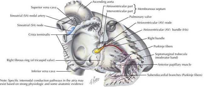
Abnormal AV conduction occurs when transmission of the normal sinus node impulses is delayed or blocked because of an abnormality in the conduction system, specifically the AV node or His-Purkinje system (Figure 45-1).
First-Degree Atrioventricular Block
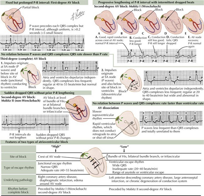
Because of an abnormal delay in conduction through the AV node, first-degree AV block results in prolongation of the PR interval above the upper limits of normal for age and heart rate (Figure 45-2). It is important to note that bradycardia does not occur because of first-degree AV block alone. This type of block can appear in otherwise healthy children as a benign phenomenon, usually related to increased vagal tone. Other causes may include cardiac surgery, rheumatic fever, Lyme disease, digoxin toxicity, and electrolyte imbalance. Isolated first-degree AV block does not require treatment unless there is progression to more advanced AV block.
Second-Degree Atrioventricular Block
Intermittent failure of AV conduction results in two forms of second-degree AV block: Mobitz type I and Mobitz type II. Each type of second-degree AV block occurs at a different level within the conduction system, thus yielding different surface ECG morphologies. Pathologic causes include myocarditis, cardiomyopathy, myocardial infarction, digitalis toxicity, congenital heart defects, and cardiac surgery.
Mobitz Type I (Wenckebach phenomenon) occurs at the level of the AV node, yielding progressive lengthening in the PR interval until one QRS complex or ventricular depolarization fails to occur (see Figure 45-2). This may occur in otherwise healthy children with parasympathetic dominance as well as in trained athletes and is frequently observed during monitoring of heart rates during sleep. After confirmation via surface ECG, management should focus on treating any underlying pathologic causes.
Mobitz type II occurs at the level of the bundle of His and is defined as the intermittent loss of AV conduction without preceding lengthening of the PR interval. This form of second-degree AV block is less common and suggests a more serious form of AV conduction disorder in which progression to complete heart block with hemodynamic compromise is more likely. Although similar causes exist between Mobitz type I and type II, consultation with a pediatric cardiologist is indicated (see Figure 45-2).
Third-Degree Atrioventricular Block or Complete Atrioventricular Block
Complete AV block is defined as complete interruption of atrial impulse propagation, resulting in atrial and ventricular activity that is independent of each other (AV dissociation). Surface ECG morphology will show regular P waves at a rate appropriate for age but with QRS complexes occurring at regular and slower rates than the atrial rate with no consistent relationship between P waves and QRS complexes. Both congenital and acquired forms of complete AV block occur and should be suspected in any patient with symptomatic bradycardia (see Figure 45-2).

Stay updated, free articles. Join our Telegram channel

Full access? Get Clinical Tree