Chapter 7 Antepartum Care
PRECONCEPTION AND PRENATAL CARE, GENETIC EVALUATION AND TERATOLOGY, AND ANTENATAL FETAL ASSESSMENT
Preconception Care
Ideally, prenatal care should begin before pregnancy. Organogenesis begins early in pregnancy, and placental development starts with implantation at 7 days postconception. Poor placental development has been linked to such pregnancy complications as preeclampsia, preterm birth, and intrauterine growth restriction and may play a role in fetal programming of chronic diseases in later life. By the time most pregnant women have their first prenatal visit, it is often too late to prevent some birth defects or defective placental development.
More importantly, early prenatal care is often too late to restore allostasis. Allostasis refers to the body’s ability to maintain stability through change. Examples include feedback inhibition on the hypothalamic-pituitary-adrenal (HPA) axis to keep the body’s stress response in check, or modulation of the body’s inflammatory response by the HPA axis. In the face of chronic and repeated stress (psychological or biologic), however, these systems can wear out. If a woman enters pregnancy with worn-out allostatic systems (e.g., dysregulated stress or inflammatory response), she may be more vulnerable to a number of pregnancy complications, including preterm birth.
The growing recognition of the limits of prenatal care and the importance of women’s health before pregnancy has drawn increasing attention to preconception care. As defined by the U.S. Centers for Disease Control and Prevention, preconception care is a set of interventions that aim to identify and modify biomedical, behavioral, and social risks to a woman’s health or pregnancy outcome through prevention and management. The American College of Obstetricians and Gynecologists (ACOG) recommends that a routine visit by any woman who may, at some time, become pregnant presents an opportunity to promote preconception health, whether or not she is planning on getting pregnant. Men should also get preconception care, though the content of preconception care for men is less well defined.
Several models of preconception care have been developed. Major components of preconception care include risk assessment, health promotion, and medical and psychosocial interventions and follow-up, as summarized in Table 7-1. There is currently no consensus on the timing of preconception care, probably because there are different ideas about what preconception care should be or do. For some, preconception care means a single prepregnancy checkup a few months before couples attempt to conceive. A single visit, however, may be too little too late to address some problems (e.g., promoting smoking cessation or healthy weight) and will miss those pregnancies that are unintended at the time of conception (about half of all pregnancies in the United States). For others, preconception care means all well-woman care, from prepubescence to menopause. In practice, however, asking providers to squeeze more into an already hurried routine visit may not be feasible, and some components (e.g., genetic screening or laboratory testing) may not be indicated for every woman at every visit.
TABLE 7-1 ELEMENTS OF PRECONCEPTION COUNSELING AND CARE
Major Components of Preconception Care | Risk Assessment |
---|---|
Reproductive life plan | Ask your patient if she plans to have any (more) children and how long she plans to wait until she (next) becomes pregnant. Help her develop a plan to achieve those goals. |
Past reproductive history | Review prior adverse pregnancy outcomes, such as fetal loss, birth defects, low birth weight, and preterm birth, and assess ongoing biobehavioral risks that could lead to recurrence in a subsequent pregnancy. |
Past medical history | Ask about past medical history such as rheumatic heart disease, thromboembolism, or autoimmune diseases that could affect future pregnancy. Screen for ongoing chronic conditions such as hypertension and diabetes. |
Medications | Review current medication use. Avoid category X drugs and most category D drugs unless potential maternal benefits outweigh fetal risks (see Box 7-1). Review use of over-the-counter medications, herbs, and supplements. |
Infections and immunizations | Screen for periodontal, urogenital, and sexually transmitted infections as indicated. Discuss TORCH (toxoplasmosis, other, rubella, cytomegalovirus, and herpes) infections and update immunization for hepatitis B, rubella, varicella, Tdap (combined tetanus, diphtheria, and pertussis), human papillomavirus, and influenza vaccines as needed. |
Genetic screening and family history | Assess risk for chromosomal or genetic disorders based on family history, ethnic background, and age. Offer cystic fibrosis screening. Discuss management of known genetic disorders (e.g., phenylketonuria, thrombophilia) before and during pregnancy. |
Nutritional assessment | Assess anthropometric (body mass index), biochemical (e.g., anemia), clinical, and dietary risks. |
Substance abuse | Ask about smoking, alcohol, drug use. Use T-ACE (tolerance, annoyed, cut down, eye opener) or CAGE (cut-down, annoyed, guilty, eye-opener) questions to screen for alcohol and substance abuse. |
Toxins and teratogens | Review exposures at home, neighborhood, and work. Review Material Safety Data Sheet and consult local Teratogen Information Service as needed. |
Psychosocial concerns | Screen for depression, anxiety, intimate-partner violence, and major psychosocial stressors. |
Physical examination | Focus on periodontal, thyroid, heart, breasts, and pelvic examination. |
Laboratory tests | Check complete blood count, urinalysis, type and screen, rubella, syphilis, hepatitis B, HIV, cervical cytology; screen for gonorrhea, chlamydia, and diabetes in selected populations. Consider thyroid-stimulating hormone. |
Major Components of Preconception Care | Health promotion |
Family planning | Promote family planning based on a woman’s reproductive life plan. For women who are not planning on getting pregnant, promote effective contraceptive use and discuss emergency contraception. |
Healthy weight and nutrition | Promote healthy prepregnancy weight through exercise and nutrition. Discuss macronutrients and micronutrients, including 5-a-day and daily intake of multivitamin containing folic acid. |
Health behaviors | Promote such health behaviors as nutrition, exercise, safe sex, effective use of contraception, dental flossing, and use of preventive health services. Discourage risk behaviors such as douching, nonuse of seat belt, smoking, and alcohol and substance abuse. |
Stress resilience | Promote healthy nutrition, exercise, sleep, and relaxation techniques; address ongoing stressors such as intimate partner violence; identify resources to help patient develop problem-solving and conflict resolution skills, positive mental health, and relational resilience. |
Healthy environments | Discuss household, neighborhood, and occupational exposures to metals, organic solvents, pesticides, endocrine disruptors, and allergens. Give practical tips such as how to reduce exposures during commuting or picking up dry cleaning. |
Preconception care is probably more than a single prepregnancy visit and less than all well-woman care. A good place to start is to ask every woman at every visit about her reproductive life plan. A reproductive life plan is a set of personal goals about having or not having children based on personal values and resources, and a plan to achieve those goals. The provider should ask the woman if she plans to have any (more) children, and how long she plans to wait until she (next) becomes pregnant. If it is within the next 1 or 2 years, the provider should bring her and her partner back for a full assessment and counseling. The schedule of follow-up visits should be individualized according to identified risks. If she does not plan on becoming pregnant in the next 1 to 2 years or ever, the provider should continue to provide well-woman care but make sure she has effective contraception if needed and update her reproductive life plan at every routine visit. Because about half of all pregnancies in the United States are unplanned, preconception counseling is recommended for every woman of reproductive age.
One example of how preconception care can improve obstetric outcomes is the opportunity to counsel and appropriately change dietary behavior. Women in the reproductive age group should be instructed to take multivitamins containing folic acid and in addition omega-3 fatty acids. Women who are underweight (body mass index [BMI] <19) have a greater risk for having a low-birth-weight or premature infant, and women who are obese (BMI >29) are at significantly greater risk for obstetric complications, including pregnancy-induced hypertension, diabetes mellitus, and fetal macrosomia. It is important that nutrition be balanced for at least 3 months before conception. Attempts at weight loss too soon before conception may have deleterious effects on fetal development.
Prenatal Care
The three basic components of prenatal care are (1) early and continuing risk assessment, (2) health promotion, and (3) medical and psychosocial interventions and follow-up. Risk assessment includes a complete history, a physical examination, laboratory tests, and assessment of fetal growth and well-being. Health promotion consists of providing information on proposed care, enhancing general knowledge of pregnancy and parenting, and promoting and supporting healthful behaviors. Interventions include treatment of any existing illness, provision of social and financial resources, and referral to and consultation with other specialized providers.
THE FIRST PRENATAL VISIT
The first prenatal visit provides an opportunity to assess or review medical, reproductive, family, genetic, nutritional, and psychosocial histories. Women whose health may be seriously jeopardized by the pregnancy, such as those with Eisenmenger’s syndrome or a history of peripartum cardiomyopathy, should be counseled about the option of terminating the pregnancy. Such reproductive histories as preterm birth, low birth weight, preeclampsia, stillbirth, congenital anomalies, and gestational diabetes are important to obtain because of the substantial risk for recurrence. Women with prior cesarean delivery should be asked about the circumstances of the delivery, and discussion about options for the mode of delivery for the current pregnancy should be initiated. Additionally, the importance of screening women for domestic violence cannot be overemphasized. As many as 20% of women are physically abused during pregnancy (most studies report a prevalence that clusters around 4% to 8%), making abuse more common than preeclampsia, diabetes, and other conditions that are routinely screened for during prenatal care.
Standardized forms have been developed to facilitate overall prenatal risk assessment. One such system is the Problem Oriented Prenatal Risk Assessment System, or POPRAS (www.POPRAS.com).
Prenatal laboratory testing should be undertaken as outlined in Table 7-1, if not done during preconception care. Screening for and treating asymptomatic bacteriuria significantly reduces the risk for pyelonephritis and preterm delivery.
Voluntary and confidential HIV counseling and testing should be offered and documented in the medical record. Diagnosis and treatment significantly reduce the risk for vertical transmission. Other tests, such as screening for sexually transmitted infections like gonorrhea and chlamydia, are generally considered routine. All pregnant women at high risk for tuberculosis should be screened with a purified protein derivative (PPD) skin test when they begin prenatal care.
Additionally, the clinician should use the first prenatal visit to confirm pregnancy and determine viability, estimate gestational age and due date, diagnose and deal with early pregnancy loss, provide genetic counseling and information about teratology, and provide advice on alleviating unpleasant symptoms during pregnancy. Information about nutrition, behavioral changes to expect, and the benefits of breastfeeding should be provided as prenatal care progresses. Clinical pelvimetry should be performed sometime before labor begins.
Confirming Pregnancy and Determining Viability
Women most commonly present to the clinician after missed menses. About 30% to 40% of all pregnant women will have some bleeding during early pregnancy (e.g., implantation bleeding), which may be mistaken for a period. Therefore, a pregnancy test should be performed in all women of reproductive age who present with abnormal vaginal bleeding.
The pregnancy test detects human chorionic gonadotropin (hCG) in the serum or the urine. The most widely used standard is the First International Reference Preparation (1st IRP). The hCG molecule is first detectable in serum 6 to 8 days after ovulation. A titer of less than 5 IU/L is considered negative, and a level above 25 IU/L is a positive result. Values between 6 and 24 IU/L are considered equivocal, and the test should be repeated in 2 days. A concentration of about 100 IU/L is reached about the date of expected menses. Most qualitative urine pregnancy tests can detect hCG above 25 IU/L.
It is important to differentiate a normal pregnancy from a nonviable or ectopic gestation. In the first 30 days of a normal gestation, the level of hCG doubles every 2.2 days. In patients whose pregnancies are destined to abort, the level of hCG rises more slowly, plateaus, or declines.
The use of transvaginal ultrasonography has improved the accuracy of predicting viability in early pregnancies. Using transvaginal ultrasonography, the gestational sac should be seen at 5 weeks of gestation or a mean hCG level of about 1500 IU/L (1st IRP). The fetal pole should be seen at 6 weeks or a mean hCG level of about 5200 IU/L. Fetal cardiac motion should be seen at 7 weeks or a mean hCG level of about 17,500 IU/L. The presence of a gestational sac of 8 mm (mean sac diameter) without a demonstrable yolk sac, 16 mm without a demonstrable embryo, or the absence of fetal cardiac motion in an embryo with a crown-rump length of greater than 5 mm indicates probable embryonic demise. When there is any doubt about these measurements, it is best to repeat the evaluation in 1 week before terminating the pregnancy.
INCIDENCE OF EARLY PREGNANCY LOSS
Because the incidence of conception is unknown, the incidence of spontaneous abortion (miscarriage) cannot be determined with certainty. Spontaneous abortion occurs in 10% to 15% of clinically recognizable pregnancies. The term biochemical pregnancy refers to the presence of hCG in the blood of a woman 7 to 10 days after ovulation but in whom menstruation occurs when expected. In other words, conception has occurred, but spontaneous loss of the gestation takes place without prolongation of the menstrual cycle. When both clinical and biochemical pregnancies are considered, evidence would suggest that more than 50% of all conceptions are lost, the majority in the 14 days following conception.
Real-time ultrasonography has been extensively used to monitor the intrauterine events of the first trimester of pregnancy. If a live, appropriately grown fetus is present at 8 weeks’ gestation, the fetal loss rate over the next 20 weeks (up to 28 weeks) is on the order of 3%.
TYPES OF SPONTANEOUS ABORTION
The terms and definitions in the remainder of this chapter refer only to clinically recognizable pregnancies.
Threatened Abortion
The term threatened abortion is used when a pregnancy is complicated by vaginal bleeding before the 20th week. Pain may not be a prominent feature of threatened abortion, although a lower abdominal dull ache sometimes accompanies the bleeding. Vaginal examination at this stage usually reveals a closed cervix. About one third of pregnant women have some degree of vaginal bleeding during the first trimester, and 25% to 50% of threatened abortions eventually result in loss of the pregnancy.
Inevitable Abortion
In a case of inevitable abortion, a clinical pregnancy is complicated by both vaginal bleeding and cramp-like lower abdominal pain. The cervix is frequently partially dilated, contributing to the inevitability of the process.
Incomplete Abortion
In addition to vaginal bleeding, cramp-like pain, and cervical dilation, an incomplete abortion involves the passage of products of conception, often described by the woman as looking like pieces of skin or liver.
Complete Abortion
In complete abortion, after passage of all the products of conception, the uterine contractions and bleeding abate, the cervix closes, and the uterus is smaller than the period of amenorrhea would suggest. In addition, the symptoms of pregnancy are no longer present, and the pregnancy test becomes negative.
Missed Abortion
The term missed abortion is used when the fetus has died but is retained in the uterus, usually for more than 6 weeks. Because coagulation problems may develop, fibrinogen levels should be checked weekly until the fetus and placenta are expelled (spontaneously) or removed surgically.
Recurrent Abortion
Three successive spontaneous abortions usually occur before a patient is considered as a recurrent aborter. Many clinicians feel that two successive first-trimester losses or a single second-trimester spontaneous abortion is justification for an evaluation of a couple for the causes of the pregnancy losses (see page 77, “Patients Who Require Genetic Counseling”).
ETIOLOGY
Although many factors may result in the loss of a single pregnancy, relatively few factors are present consistently in couples who abort recurrently. Cause-and-effect relationships in individual patients are frequently difficult to determine.
General Maternal Factors
Infection with Mycoplasma, Listeria, or Toxoplasma should be specifically sought in women with recurrent abortions because despite being found infrequently, they are all treatable with antibiotics. Maternal smoking and alcohol consumption are associated with an increased incidence of chromosomally normal abortions. Women who smoke 20 or more cigarettes daily and consume more than seven standard alcoholic drinks per week have a fourfold increased risk for spontaneous abortion. There is a doubling of the risk for spontaneous abortion with as few as two drinks a week.
There is very little evidence that a sudden physical or emotional shock can cause pregnancy loss, but psychodynamic factors may contribute to recurrent abortion in a few cases.
Three medical disorders are commonly linked to spontaneous abortion: (1) diabetes mellitus, (2) hypothyroidism, and (3) systemic lupus erythematosus (SLE). The evidence linking diabetes mellitus with spontaneous abortion is not conclusive, and severe hypothyroidism is more often associated with disordered ovulation than spontaneous abortion. Up to 40% of clinical pregnancies are lost in women with SLE, and such patients have an increased risk for pregnancy loss before developing the clinical stigmata of the disease (see Chapter 16).
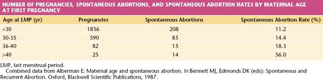
The risk for abortion increases with maternal age (Table 7-2). If a live fetus is demonstrated by ultrasonography at 8 weeks’ gestational age, however, fewer than 2% will abort spontaneously when the mother is younger than 30 years of age. If she is older than 40 years, the risk exceeds 10%, and it may be as high as 50% at age 45 years. The probable explanation is the increased incidence of chromosomally abnormal conceptus in older women.
Local Maternal Factors
No prospective study has been able to demonstrate unequivocally that a normal pregnancy can be lost as a result of abnormal hormone production by either the corpus luteum or the placenta. In addition to this, no controlled trial of exogenous hormones has been able to demonstrate any benefit, and there is some evidence that exogenous sex steroids may indeed be teratogenic.
Uterine abnormalities, including cervical incompetence, congenital abnormalities of the uterine fundus (as may result from gestational exposure to diethylstilbestrol), and acquired abnormalities of the uterine fundus are known to be associated with pregnancy loss.
Cervical incompetence occurs under a number of circumstances. The incompetence is usually the result of trauma. This occurs most frequently from mechanical dilation of the cervix at the time of termination of pregnancy, but it may also occur at the time of curettage. The diagnosis of cervical incompetence is usually made when a mid-trimester pregnancy is lost with a clinical picture of sudden unexpected rupture of the membranes, followed by painless expulsion of the products of conception.
There continues to be controversy surrounding cervical incompetence, with some experts suggesting that cervical incompetence is, in most instances, a variant of preterm delivery, occurring at a time when there is an associated finding of asymptomatic ascending infection.
When cervical incompetence is suspected during pregnancy (e.g., history of cervical incompetence in a previous pregnancy or of cone biopsy of the cervix), sequential ultrasonography of the cervix and lower uterine segment may identify the problem before a pregnancy loss occurs.
A congenitally abnormal uterus may be associated with pregnancy loss in both the first and second trimesters. Surgical correction of the abnormality, particularly with a history of second-trimester loss, is frequently successful. The diagnosis of these abnormalities is made by either hysterography or hysteroscopy. Complete evaluation of the congenitally abnormal uterus usually requires laparoscopic, hysteroscopic, and hysterographic examination before any management plan can be made.
The most commonly acquired abnormalities of the uterus with the potential to affect fecundity are submucous fibroids. Although these tend to occur more frequently in women in their late 30s, they should be considered when investigating pregnancy loss in all women. Removal of submucous fibroids and large (>6 cm) intramural ones is associated with improved fecundity, especially when they distort the endometrial cavity. Subserous fibroids do not appear to affect fecundity.
Intrauterine adhesions result from trauma to the basal layer of the endometrium from previous surgery or infection. When most of the uterine cavity has been obliterated (Asherman’s syndrome), amenorrhea results; but much more frequently, fewer intrauterine adhesions (synechiae) are present with reasonably normal menses, and these lesions are not even suspected until a pregnancy is attempted and lost. Surgical correction of these intrauterine adhesions is recommended to improve fecundity.
Fetal Factors
The most common cause of spontaneous abortion is a significant genetic abnormality of the conceptus. In spontaneous first-trimester abortions, about two thirds of fetuses have significant chromosomal anomalies, with about half of these being autosomal trisomies and most of the remainder being triploid, tetraploid, or 45 X monosomies. Fortunately, most of these are not inherited from either mother or father and are single nonrecurring events. When seen on ultrasonography before spontaneous abortion occurs, many such pregnancies appear to consist of an empty gestational sac. When a fetus is present in many late first-trimester and early second-trimester abortions, it is often significantly abnormal, either genetically or morphologically. It seems that nature has a way of identifying some of its major mistakes and causing them to abort.
Chromosomal Factors
Occasionally, fetal chromosomal abnormalities occur as a result of a chromosomal rearrangement (balanced translocation, inversion) in either parent. Therefore, karyotyping is important for evaluation of couples suffering from recurrent abortion.
Immunologic Factors
A successful pregnancy depends on a number of immunologic factors that allow the host (mother) to retain an antigenically foreign product (fetus) without rejection taking place (see Chapter 6). The precise mechanism of this immunologic anomaly is not fully understood, but the immunologic functioning of some women, particularly those who abort recurrently, is different from that of women who carry pregnancies to term. The immunologic relationship between male and female in such a couple may be regarded as abnormal, and in some instances, treatment of this condition may result in a successful pregnancy.
MANAGEMENT
Threatened Abortion
A threatened abortion is best managed by an ultrasonic examination to determine whether the fetus is present and, if so, whether it is alive. Of those in whom a live fetus is present, 94% will produce a live baby, although the incidence of preterm delivery in these cases may be somewhat higher than in those who do not bleed in the first trimester. Once a live fetus has been demonstrated to the couple on ultrasonography, management consists essentially of reassurance; however, they should be encouraged to undergo first trimester screening for chromosome abnormalities such as trisomy 13, 18, or 21. There is no need for admission to hospital nor is there any evidence that bed rest improves the prognosis.
Incomplete Abortion
Until bleeding has stopped or is minimal, it is best to insert an intravenous line and take blood for grouping and crossmatching because shock may occur from hemorrhage or sepsis. Once the patient’s condition is stable, the remaining products of conception should be evacuated from the uterus under appropriate pain control. These tissues should be sent for pathologic evaluation. An incomplete abortion that is infected must be managed vigorously. Delay in treatment may result in overwhelming sepsis that may lead to renal and hepatic failure, disseminated intravascular coagulation (DIC), and even death.
Missed Abortion
Suspected missed abortion should be confirmed by ultrasound. Once the diagnosis has been made, it is appropriate to evacuate the retained products of conception surgically to minimize the risk for sepsis and DIC and to reduce the extent of hemorrhage and the degree of pain that accompanies the spontaneous expulsive process.
General Management Considerations
When the patient is Rh negative and does not have Rh (anti-D) antibodies, prophylactic Rho(D) immune globulin (Rho-GAM) should be administered. All couples who have had a pregnancy loss should be seen and counseled some weeks after the event. At this time, questions that the couple may have can be answered, the findings of any pathologic studies discussed, and reassurance given about their chances of reproductive success in the future.
Recurrent Abortion
As far as the mother is concerned, it is appropriate to rule out the presence of systemic disorders such as diabetes mellitus, SLE, and thyroid disease, and it is also necessary to test for the presence of a lupus anticoagulant. Paternal and maternal chromosomes should be evaluated, and hysteroscopy or hysterography should be performed to evaluate the uterine cavity. Given the possibility of the pregnancy losses being caused by infectious agents, it is also appropriate to rule out the presence of Mycoplasma, Listeria, Toxoplasma, Treponema, cytomegalovirus, and Brucella.
More than half of couples with recurrent losses will have normal findings during an evaluation. When a specific etiologic factor is found, appropriate management often leads to reproductive success. Many of the congenital abnormalities of the uterus can now be diagnosed using pelvic ultrasonography and may no longer require laparotomy for repair. Cervical incompetence is managed by the placement of a cervical suture (cerclage) at the level of the internal os, and this suture is best placed in the first trimester, after a live fetus has been demonstrated on ultrasonography. The effectiveness of prophylactic cervical cerclage (see Chapters 17 and 19) in preventing recurrent loss from cervical incompetence has not been conclusively established.
Estimating Gestational Age and Date of Confinement
Gestational age should be determined during the first prenatal visit. Accurate determination of gestational age may become important later in pregnancy for the management of obstetric conditions such as preterm labor, intrauterine growth restriction, and postdate pregnancy. Clinical assessment to determine gestational age is usually appropriate for the woman with regular menstrual cycles and a known last menstrual period that was confirmed by an early examination. Estimated date of confinement (EDC) or “due date” may be determined by adding 9 months and 7 days to the first day of the last menstrual period.
Ultrasonography may also be used to estimate gestational age. Measurement of fetal crown-rump length between 6 and 11 weeks of gestation can define gestational age to within 7 days. At 12 to 20 weeks, gestational age can be determined within 10 days by the average of multiple measurements (e.g., biparietal diameter, femur length, abdominal and head circumferences). Thereafter, measurements become less reliable with advancing gestation (±3 weeks in the third trimester).

Stay updated, free articles. Join our Telegram channel

Full access? Get Clinical Tree

