Chapter 32 Amenorrhea, Oligomenorrhea, and Hyperandrogenic Disorders∗
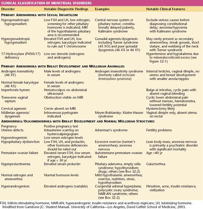
Amenorrhea, or the absence of menses, is a common symptom of several pathophysiologic states. This condition traditionally has been divided into primary amenorrhea, in which menarche (the first menses) has not occurred, and secondary amenorrhea, in which menses has been absent for 6 months or more. A more functional or clinical division of menstrual disorders based on initial history and physical examination would be as follows: primary amenorrhea with sexual infantilism, primary amenorrhea with breast development and müllerian anomalies, and amenorrhea and oligomenorrhea with breast development and normal müllerian structures. The last group includes disorders causing primary as well as secondary amenorrhea, oligomenorrhea, and the hyperandrogenic states (Table 32-1).
Primary Amenorrhea
The diagnosis of primary amenorrhea is made when no spontaneous uterine bleeding has occurred by the age of 16 years. The workup should be initiated earlier if there is no evidence of breast development (thelarche) by age 14 years or if the patient has failed to menstruate (menarche) spontaneously within 2 years of thelarche. The presence of normal breast development confirms gonadal secretion of estrogen but not necessarily the presence of ovarian tissue. The presence of normal amounts of pubic and axillary hair confirms gonadal or adrenal secretion of androgens as well as the presence of functional androgen receptors.
PRIMARY AMENORRHEA WITH SEXUAL INFANTILISM
Patients with primary amenorrhea and no secondary sexual characteristics (sexual infantilism) display the absence of gonadal hormone secretion. The differential diagnosis is based on whether the defect is the result of a lack of gonadotropin secretion (hypogonadotropic hypogonadism) or an inability of the ovaries to respond to gonadotropin (hypergonadotropic hypogonadism due to gonadal agenesis or dysgenesis). The distinction can be made by the measurement of a basal serum follicle-stimulating hormone (FSH).
Hypogonadotropic Primary Amenorrhea and Sexual Infantilism
Patients with hypogonadotropic hypogonadism have low FSH levels, whereas patients with hypergonadotropic hypogonadism (e.g., gonadal dysgenesis) have elevated FSH levels in the menopausal range (>20 or 40 mIU/L, depending on the assay used). The measurement of serum luteinizing hormone (LH) is of limited additional diagnostic value. The absence of breast development is indicative of inadequate secretion of estrogen.
Hypogonadotropic hypogonadism may be caused by lesions of the hypothalamus or pituitary gland or by functional disorders that result in inadequate gonadotropin-releasing hormone (GnRH) synthesis and release. Because patients with sexual infantilism caused by hypogonadotropic hypogonadism may have a craniopharyngioma or other central nervous system tumor, magnetic resonance imaging (MRI) or computerized tomography (CT) of the hypothalamic-pituitary area is recommended.
Hypogonadotropic hypogonadism resulting in primary amenorrhea and sexual infantilism may also be the result of lesions of the pituitary, including prolactin-secreting adenomas, or a general process of pituitary failure. These patients should be screened for other pituitary hormonal deficiencies by testing for thyroid-stimulating hormone (TSH), growth hormone, and adrenocorticotropic hormone (ACTH).
Finally, apparent hypogonadotropic hypogonadism may actually represent constitutionally delayed puberty. This delay in the normal onset of puberty is generally attributed to undefined hereditary factors because there is commonly a history of late puberty in family members. Constitutional delay of puberty is a diagnosis of exclusion.
Hypergonadotropic Primary Amenorrhea and Sexual Infantilism
Patients with hypergonadotropic hypogonadism have some form of failed gonadal development or premature gonadal failure and will have elevated FSH levels. These patients may have gonadal agenesis (the absence or early disappearance of the normal gonad). Examples in males who may appear to be female in some cases are pure gonadal dysgenesis, or the testicular regression syndrome. These patients have an apparently normal 46 XY karyotype but lack testicular development. If fetal testicular regression occurs between 8 and 10 weeks of gestation, they may have female external genitalia with or without ambiguity in addition to a lack of gonads, a hypoplastic uterus (secondary to absent secretion of antimüllerian hormone), and rudimentary genital ducts (Swyer syndrome). Regression of the testes after 12 to 14 weeks results in variable development of male external genitalia. Anorchia, or streak gonads, occurs with testicular regression syndrome.
Other individuals with hypergonadotropic primary amenorrhea and sexual infantilism may have gonadal dysgenesis, or the presence of an abnormally developed gonad due to chromosomal defects. The differential diagnosis includes 45 XO (Turner syndrome), a structurally abnormal X chromosome, mosaicism with or without a Y chromosome, and pure gonadal dysgenesis (46 XX and 46 XY). Although most affected patients show no signs of secondary sexual characteristics, occasionally an individual with mosaicism or Turner syndrome will have sufficient ovarian follicular activity and secrete enough estrogen to cause breast development, menstruation, ovulation, and rarely even pregnancy.
In individuals with the presence of a Y chromosome, there is a risk for developing a gonadoblastoma (a benign germ cell tumor of the gonad) and eventually dysgerminoma (a malignant germ cell tumor). All patients with hypergonadotropic hypogonadism should have a karyotype performed. Because it is important to identify mosaicism, a greater number of white blood cells (>35) should be karyotyped.
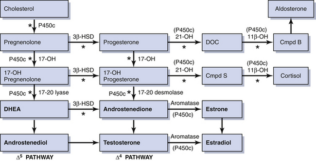
Rarely, some patients with primary amenorrhea and sexual infantilism have a defect of estrogen and androgen production. One example of this is a 17-hydroxylase (P450c17) deficiency, which prevents the synthesis of these sex steroids (Figure 32-1). These individuals have hypertension and hypokalemia caused by mineralocorticoid excess. Other patients, such as those with a 46 XY karyotype and Leydig cell agenesis, may lack the cells necessary for sex steroid production. Because the Leydig cells in the testicle are responsible for producing testosterone, these individuals are born with female external genitalia.

FIGURE 32-1 Diagrammatic representation of the steroid biosynthetic pathways. The asterisk refers to specific enzyme defects that result in congenital adrenal hyperplasia. Cmpd B, corticosterone; Cmpd S, II-deoxycortisol; DHEA, dehydroepiandrosterone; DOC, desoxycorticosterone; HSD, hydroxysteroid dehydrogenase; OH, hydroxylase; P450c, cytochrome P450.
Patients with sexual infantilism may be treated to stimulate breast development by very gradually increasing estrogen doses. One commonly used regimen is to start with 0.3 mg conjugated estrogen every other day and slowly increase over 3- to 6-month intervals. This treatment should be guided by the presence or absence of mastalgia and the rate of breast development. The estrogen can be safely increased to 0.6 mg or more daily if necessary.
Individuals with persistent hypogonadotropic hypogonadism who seek fertility require either human menopausal gonadotropin injections or pulsatile GnRH administered by an infusion pump. Patients with gonadal dysgenesis and 17-hydroxylase deficiency who have a normal uterus and cervix can achieve pregnancy only by in vitro fertilization using donor oocytes.
PRIMARY AMENORRHEA WITH BREAST DEVELOPMENT AND MÜLLERIAN ANOMALIES
Patients with primary amenorrhea, breast development, and some defect of müllerian structures fall into two categories: those with complete androgen insensitivity syndrome (AIS), formerly called testicular feminization, and those with müllerian dysgenesis or agenesis. The distinction between these two diagnoses can be made by the measurement of a serum testosterone level and determination of the karyotype.
Androgen Insensitivity Syndrome
Patients with complete androgen insensitivity syndrome have a defect in the androgen receptor. Their karyotype is 46 XY, and they demonstrate male levels of testosterone, although usually on the lower side of normal. They may also have mildly elevated FSH and LH levels because their testes are located within the abdominal wall or cavity (cryptorchic). This location, with greater body heat, typically does not allow for normal male hormonal secretion. Breast development (with smaller nipples and areolae than normal genotypical females) is caused by the testicular secretion of estrogens and by the conversion of circulating androgen to estrogens in the liver and elsewhere. The testicles of individuals with AIS secrete normal male amounts of antimüllerian hormone; therefore, patients have only a vaginal dimple and no uterus. Treatment should consist of gonadal resection to avoid neoplasia (i.e., gonadoblastomas and dysgerminomas) once puberty is complete. The creation of a neovagina when the patient is prepared for sexual activity is possible by surgical and nonsurgical methods. Psychological counseling is an important component in the care of these patients.
Müllerian Dysgenesis or Agenesis
Patients with primary amenorrhea, breast development, and a 46 XX karyotype have levels of testosterone appropriate for females. This clinical diagnosis may be caused by müllerian defects that cause obstruction of the vaginal canal (e.g., imperforate hymen or a transverse vaginal septum) or by the absence of a normal cervix or uterus and normal fallopian tubes. An imperforate hymen should be suspected in adolescents who report monthly dysmenorrhea in the absence of vaginal bleeding (see Figure 18-7, pg 237). Clinically, these patients often present with a vaginal bulge and a midline cystic mass on rectal examination. Ultrasonography confirms the presence of a normal uterus and ovaries with a hematocolpos. These patients should be treated with hymenectomy.
Alternatively, women may present with similar symptoms but without a vaginal bulge. When ultrasonography confirms a normal uterus and ovaries, a transverse, obstructing vaginal septum (see Figure 18-8, pg 237) or cervical agenesis should be suspected. MRI is the diagnostic procedure of choice in these patients. If the MRI scan confirms a transverse septum, surgical correction is indicated. Surgical construction of a functional cervix is extremely difficult. In general, it is recommended that these women undergo hysterectomy.
Creation of a neovagina can be accomplished using one of two general approaches. The Frank method of vaginal dilation uses dilation of the vaginal pouch with vaginal forms (usually thermoplastic acrylic resin [Lucite] dilators) over the course of weeks to months. Alternatively, a McIndoe vaginoplasty, which involves the surgical creation of a neovaginal space using a split-thickness skin graft, may be performed. Both of these methods should be initiated and performed close to the time when the patient anticipates having vaginal intercourse.
Congenital anatomic abnormalities of the uterus or vagina, or both, are often associated with renal abnormalities such as a unilateral solitary kidney or a double renal collecting system, among others. Therefore, these patients should have an intravenous pyelogram or other diagnostic study to confirm a normal urinary system.
Amenorrhea and Oligomenorrhea with Breast Development and Normal Müllerian Structures
Disorders in which the patient has breast development and a demonstrable cervix and uterine fundus on physical examination may cause primary as well as secondary amenorrhea, or may present as oligomenorrhea (menstrual cycles at greater than 35- to 45-day intervals).
All patients with menstrual bleeding disorders should be tested for pregnancy. Once pregnancy has been excluded, these individuals can be characterized as shown in Table 32-1. Initial history taking should include questions about the timing of thelarche, pubarche, and menarche. The timing and development of the menstrual disorder (present since puberty or new), significant weight change, strenuous exercise activities, dietary habits, sexual activity, concomitant illnesses or complaints, abnormal facial or body hair growth, scalp hair loss, acne, and the presence or absence of hot flashes and vaginal dryness should be noted. A comprehensive list of medications and dietary supplements taken should be obtained.
UTERINE DEFECTS
Women who do not have withdrawal bleeding after a hormonal challenge test and who have a history of uterine instrumentation, particularly a dilation and curettage, following vaginal delivery or pregnancy termination may have Asherman’s syndrome. This interesting syndrome is characterized by intrauterine scarring (synechiae), and these patients may have normal ovulatory cycles with cyclic premenstrual symptoms. Patients with Asherman’s syndrome should be evaluated by hysterosalpingography or sonohysterography. Hysteroscopic treatment with excision of the synechiae and normalization of the uterine cavity is the treatment of choice.
AMENORRHEA AND OLIGOMENORRHEA ASSOCIATED WITH HYPOESTROGENISM
The differential diagnosis for patients with amenorrhea associated with low levels of estrogen includes hypothalamic-pituitary dysfunction (hypothalamic amenorrhea), premature ovarian failure, and hyperprolactinemia. Women in the first group have low FSH and prolactin levels, women in the second group have high FSH and normal prolactin levels, and women in the third group have high prolactin and low FSH levels.
Hypothalamic-Pituitary Dysfunction
Patients with hypothalamic amenorrhea include women experiencing severe weight loss, women undergoing excessive exercise resulting in low body fat, and women experiencing severe psychological stress. Also included are women with physical wasting from severe systemic diseases such as disseminated malignancies and patients with pituitary or central nervous system lesions. In its most severe and life-threatening form, women may have pituitary failure or anorexia nervosa. All patients with hypogonadotropic hypogonadism and hypothalamic-pituitary dysfunction should be evaluated for the status of the other pituitary hormones. Evaluation should also include an MRI of the hypothalamus and pituitary gland to exclude neoplastic and other lesions if it is uncertain whether the patient has one of the functional disorders described previously.
When hypothalamic-pituitary dysfunction cannot be resolved by identifying a modifiable underlying cause (e.g., excessive exercise), estrogen and progestin therapy, usually in the form of a combined oral contraceptive pill, is prescribed to reduce the risk for osteoporosis. This therapy is also recommended to maintain normal vaginal and breast development. In patients with anorexia nervosa, ovarian hormone therapy without weight gain will not totally prevent osteoporosis.
Premature Ovarian Failure
Premature ovarian failure is defined as ovarian failure before the age of 40 years (see Chapter 35). When it occurs in patients younger than 30 years of age, ovarian failure may be caused by a chromosomal disorder. A karyotype is performed to exclude mosaicism (i.e., some cells bearing a Y chromosome). If cells with a Y chromosome are present, a gonadectomy to prevent malignant transformation is indicated.

Stay updated, free articles. Join our Telegram channel

Full access? Get Clinical Tree

