Fig. 8.1
Histological findings of adenomyosis with foci of endometrial glands and stroma found deep within the myometrium
While the exact etiology of adenomyosis is unknown, several risk factors have been identified in clinical series. Seventy to eighty percent of adenomyosis is reported in women in their fourth and fifth decades, but it is unclear if age is indeed a risk factor or a confounding factor (Azzi 1989). The higher prevalence in older women may be due to higher rates of hysterectomy later in life. It may, however, also be due to longer exposure to hormones that over time may stimulate endometriotic glands to invaginate into the myometrial wall (Garcia and Isaacson 2011).
Multiparous women also have higher rates of adenomyosis (Bergholt et al. 2001; Levgur et al. 2005; Parazzini et al. 1997; Vercellini et al. 1995; Taran et al. 2010). The frequency of adenomyosis has been linked directly to the number of pregnancies (Vercellini et al. 1995). The invasive nature of the trophoblast into the myometrium at the time of implantation may weaken the myometrium to allow active endometrial tissue to grow into the injured lining (Ferenczy 1998; Garcia and Isaacson 2011). In addition, the increase in hormones during pregnancy may also help stimulate the invagination of endometrial implants (Levgur et al. 2005).
Pelvic surgery, like pregnancy, can also weaken the myometrium through trauma with instrumentation during dilation and curettage, transmural surgery, or cesarean section (Ferenczy 1998). However, it remains unclear if prior uterine surgery is a risk factor since only a small series of cases show that pregnancy termination and dilation and curettage have an association with adenomyosis, while other studies show no statistical relationship between this disorder with cesarean section or myomectomy (Bergholt et al. 2001; Harris and Daniel 1985; Levgur et al. 2005; Parazzini et al. 1997; Taran et al. 2010).
8.2 Clinical Manifestations and Pathophysiology
Not all women with adenomyotic uteri are symptomatic but up to 75 % of women present with either menorrhagia, dysmenorrhea, or both (Benson and Sneeden 1958; Bergholt et al. 2001). Menorrhagia is likely caused by adenomyotic foci interfering with the normal musculature of the myometrium, preventing adequate contractions and permitting greater blood loss (Ferenczy 1998; Azzi 1989). Dysmenorrhea is likely a result of uterine irritability stimulated from the increase blood and from edema of the adenomyotic foci within the myometrium (Azzi 1989). Symptoms of chronic pelvic pain and dyspareunia are also common in women with adenomyosis. The pathophysiology of these symptoms is unclear. There is, however, high prevalence of adenomyosis in 70 % of women with endometriosis, which may contribute to these symptoms (Kunz et al. 2005). It is unclear if adenomyosis plays a role in reproductive outcomes, but some studies hypothesize that adenomyotic foci may interfere with implantation or impede sperm function through immune responses causing infertility (Devlieger et al. 2003; Ota et al. 1998).
8.3 Diagnostic Procedures
As described earlier (e.g. see Sect. 8.1), the diagnosis of adenomyosis is usually made from tissue specimens collected at the time of hysterectomy. Currently, the development of high quality, noninvasive imaging modalities has also been proven to be accurate in the diagnosis of adenomyosis as well. Findings of myometrial cysts, heterogeneous myometrial echotexture, and the visualization of the endometrial–myometrial interface (known as the junctional zone) have assisted with the differential diagnosis of adenomyosis using transvaginal ultrasound and magnetic resonance imaging (Bazot et al. 2001; Dueholm and Lundorf 2007; Felde et al. 1992; Reinhold et al. 1995) (Figs. 8.2 and 8.3).
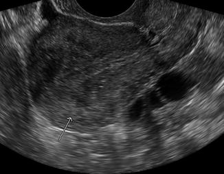
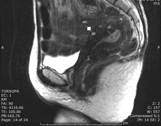
Fig. 8.2
Sagittal transvaginal sonography shows an asymmetric uterus with thickened posterior myometrial wall. The “Arrow show decreased echogencity and heterogeneity consistent with diffuse adenomyosis in the posterior wall of the uterus”
Fig. 8.3
Sagittal T2 weighted MRI images demonstrate ill defined 6.0 × 6.8 cm heterogeneous, hyperintense foci extending from the junctional zone (JZ) anteriorly measuring 21.94 mm (greater than 12 mm) suggesting adenomyosis. Myometrial cysts can be seen anteriorly (MC)
In addition, advances in hysteroscopy and laparoscopy have provided minimally invasive techniques to assist in the diagnosis of adenomyosis. Hysteroscopy provides direct visualization into the uterine cavity and allows for the option of superficial myometrial biopsies for a histological diagnosis. Hysteroscopic findings of intramyometrial lacunae, an irregular endometrium with pitting defects, or cystic hemorrhagic lesions are all suggestive of adenomyosis (Fernandez et al. 2007; Molinas and Campo 2006) (Fig. 8.4). When adenomyosis cannot be seen, hysteroscopic myometrial biopsies can be performed at the posterior wall, where adenomyosis is most frequently found (Benson and Sneeden 1958; Emge 1962). In one study, hysteroscopic myometrial biopsies were performed using a 5 mm loop electrode in fifty women with normal appearing cavities as well as a history of menorrhagia. Of those 50 women, 33 (66 %) had adenomyosis present at a depth greater than 1 mm (McCausland 1992). However, the sensitivity of the biopsy depends on extent, depth, and location of disease (Garcia and Isaacson 2011). Laparoscopic myometrial biopsies can be performed as well but have a high risk of bleeding (Popp et al. 1993).
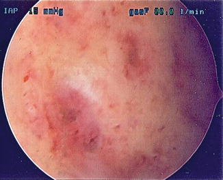
Fig. 8.4
Hysteroscopic image of adenomyosis with pathognomonic signs of pitting endometrial defects, cystic hemorrhagic lesions
8.4 Treatment
Suppressive hormonal treatments are effective for reducing symptoms of menorrhagia and dysmenorrhea but these medications only temporarily induce regression of adenomyosis during the course of therapy. Continuous oral contraceptive pills, high dose progestins, the levonogestrol intrauterine device, danazol, and GnRH agonists have all been used as conservative treatment options, as an alternative for hysterectomy. The standard of treatment still remains hysterectomy for those women who have completed child bearing. As imaging and minimally invasive diagnostic methods have evolved, less aggressive surgical options have also been developed in place of hysterectomy (Levgur 2007). Interventional radiology techniques such as uterine artery embolization and magnetic resonance guided focused ultrasound surgery have been used to treat adenomyosis (Kim et al. 2007; Rabinovici et al. 2006). Other conservative surgical treatment options such as endometrial ablation and resection, adenomyotic muscle excision and reduction, and electrocoagulation have also been performed and will be discussed in further detail.
When considering surgery as a treatment option, it is important to try to determine the area and extent of disease (Farquhar and Bronsens 2006). Because adenomyosis tends to be a diffuse process with ectopic endometrium invading throughout the entire myometrium, hysterectomy still remains the gold standard of treatment. It is the only guaranteed treatment for adenomyosis. Less invasive approaches with vaginal and laparoscopic hysterectomy are preferable to abdominal hysterectomy because of lower morbidity and faster recovery. While vaginal hysterectomy is more cost effective than the laparoscopic approach, laparoscopy may be more beneficial to the patient.
Furuhashi et al. (1998) showed that women with adenomyosis had higher rates of bladder injury at the time of vaginal hysterectomy. The authors concluded that the reason for increased rate of injury was unknown but it was hypothesized that there may be greater difficulty in identifying the supravaginal septum and the vesicovaginal or vesicocervical planes without direct visualization. In addition to allowing direct visualization for dissection, laparoscopy has the advantage of detecting and removing endometriosis that is commonly found in patients with adenomyosis (Wood 1998). The laparoscopic approach may also be beneficial due to less postoperative pain in comparison to vaginal hysterectomy (Candiani et al. 2009; Ghezzi et al. 2010). A review of 70 articles regarding hysterectomy showed lower costs and shorter operating times with vaginal hysterectomy, but the rates of blood transfusions, unexplained fever, and bleeding were higher with vaginal rather than the laparoscopic approach (Wood et al. 1997).
Adenomyosis can also appear in focal forms of circumscribed nodular aggregates of smooth muscle, endometrial glands, and stroma called adenomyomas. When adenomyomas can be localized with ultrasound or MRI, attempts can be made to excise the lesion alone. This conservative surgical option is usually attempted in women who wish to maintain fertility. However, unlike myomectomies, it can be difficult to define margins and expose lesions, which can lead to postoperative sequale affecting fertility. With excision, scar formation can affect the gestational capability of uterus, putting women at higher risk for spontaneous miscarriages (38.8 %) and uterine rupture (Wood 1998; Wang et al. 2009). However, a small study showed that conservative treatment with adenomyoma excision (mean size of 55 mm) resulted in a 70 % pregnancy rate (49/71 patients) with relief of symptoms of menorrhagia and dysmenorrheal (Wang et al. 2009). In addition, with an excisional procedure, lesions can be left behind causing patients to remain symptomatic or relapse due to the inability to clearly exposure margins in adenomyomas. Due to the low efficacy of excision of 50 %, medical therapy is often used in combination to help prevent relapses (Wang et al. 2009).
Stay updated, free articles. Join our Telegram channel

Full access? Get Clinical Tree


