65 Acute and Chronic Renal Failure
Acute Renal Failure
Acute kidney injury (AKI), previously referred to as acute renal failure (ARF), is defined as an abrupt reduction in kidney function measured by a rapid decline in glomerular filtration rate (GFR). AKI implies that an acute decline in kidney function is secondary to an injury that leads to functional or structural changes in the kidney. AKI is characterized by a disturbance of renal physiologic functions, including impairment of nitrogenous waste product excretion and inability to regulate water, electrolyte, and acid–base homeostasis. The precise incidence and prevalence of AKI in children is difficult to ascertain. The overall incidence of AKI appears to be rising because of advances in pediatric medical technology including bone marrow, hepatic, and cardiac transplantation, in surgery for congenital heart disease, and in the care of very low birth weight infants.
Etiology and Pathogenesis
The causes of AKI can be related to any process that interferes with the structure or function of the renal vasculature, glomeruli, renal tubules, interstitium, or urinary tract. The causes of AKI can be categorized as prerenal, renal (intrinsic renal disorder), or postrenal.
Prerenal
AKI results from either volume depletion caused by bleeding, gastrointestinal (vomiting, diarrhea), urinary (diuretics, diabetes insipidus), cutaneous losses (burns) or decreased effective blood volume (heart failure, cardiac tamponade, hepatorenal syndrome, shock, sepsis). In this type of AKI, the kidneys are intrinsically normal, and AKI is reversible after renal blood flow is restored by correcting the underlying disturbance. However, prolonged prerenal injury results in intrinsic renal AKI.
Renal
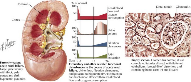
AKI (intrinsic renal disease) is the result of disorders that involve the renal vascular, glomerular, or tubular–interstitial pathology. Acute tubular necrosis (ATN) results from ischemia caused by decreased renal perfusion or injury from tubular nephrotoxins (Figure 65-1). All causes of prerenal AKI can progress to ATN if renal perfusion is not restored or nephrotoxins are not withdrawn. Nephrotoxic AKI is mostly caused by toxic tubular injury by medications, including aminoglycosides, contrast agents, amphotericin B, chemotherapeutic agents (ifosfamide, cisplatin), and acyclovir. Toxic tubular injury can also be induced by the release of heme pigments, as it occurs from myoglobinuria caused by rhabdomyolysis and hemoglobinuria caused by intravascular hemolysis. Uric acid nephropathy and tumor lysis syndrome are causes of AKI in children with leukemia. During chemotherapy, a rapid breakdown of tumor cells causes increased release and subsequent excretion of uric acid, resulting in precipitation of uric acid crystals in the tubules and renal microvasculature. Hyperphosphatemia in tumor lysis syndrome results in precipitation of calcium phosphate crystals in the tubules. Acute interstitial nephritis most commonly results from hypersensitivity reactions to drugs, including penicillin analogs (e.g., methicillin), cimetidine, sulfonamides, rifampin, nonsteroidal antiinflammatory drugs, and proton pump inhibitors, but can also be idiopathic. Glomerulonephritis of any etiology (including those caused by vasculitis, systemic lupus erythematosus, or Goodpasture’s syndrome) may present with AKI, with postinfectious glomerulonephritis being the most common cause of AKI in this group. Rapidly progressive glomerulonephritis presents as the most severe degree of any form of glomerulonephritis and presents with AKI. Vascular causes of AKI include cortical necrosis (mostly caused by hypoxic or ischemic injury in newborns), renal artery or vein thrombosis, and hemolytic-uremic syndrome (HUS).
Postrenal
AKI is caused by bilateral urinary tract obstruction unless there is a solitary kidney or caused by urethral obstruction. Examples of congenital disorders causing obstruction and AKI are posterior urethral valves (PUVs), bilateral ureteropelvic junction obstruction, and bilateral ureteroceles. Examples of acquired causes of obstruction and AKI are kidney stones and tumors.
Clinical Presentation
A careful history and physical examination can frequently identify disease processes that underlie AKI and suggest an underlying diagnosis.
Evaluation and Diagnosis
In addition to a careful history and physical examination, the initial evaluation includes additional laboratory studies.
Serum Creatinine Concentration, Acute Kidney Injury Biomarkers, and Classification of Acute Kidney Injury
AKI is diagnosed by an increase in serum creatinine concentration, which strongly suggests a decrease in the GFR. However, it is accepted that the serum creatinine concentration is an insensitive and delayed measure of decreased kidney function after AKI. Biomarkers that allow recognition of the early stages of AKI have been identified. Clinical use of these markers may permit initiation of timely therapy. These biomarkers are serum neutrophil gelatinase-associated lipocalin (NGAL) and cystatin C, urinary NGAL, interlukin-18, and kidney injury molecule-1 (KIM-1). A standardized classification of AKI has recently been proposed in adults (RIFLE criteria) and adapted for children (pediatric [p] RIFLE criteria). pRIFLE stands for risk for renal dysfunction, injury to the kidney, failure of kidney function, loss of kidney function, end-stage renal disease. These criteria better characterize AKI and reflect the course of AKI in children admitted to the intensive care unit (ICU).
Urinalysis
The urinalysis is the most important noninvasive test in the diagnostic evaluation because characteristic findings on microscopic examination of the urine sediment strongly suggest certain diagnoses. A normal or near-normal urinalysis result, characterized by few cells with little or no casts or proteinuria, suggests prerenal disease or urinary tract obstruction. Muddy brown granular casts and epithelial cell casts are highly suggestive of ATN. The finding of red blood cell (RBC) casts is diagnostic of glomerulonephritis, and heavy proteinuria is indicative of glomerular disease, both suggesting AKI caused by glomerulonephritis. The presence of many white blood cells (pyuria) suggests urinary tract infection, and white blood cells with granular or waxy casts and mild to moderate proteinuria suggest tubular or interstitial disease. Eosinophils in the urine suggest interstitial nephritis caused by a hypersensitivity reaction. Hematuria can be seen in glomerulonephritis and renal vein thrombosis and with renal calculi.
Urine Sodium Excretion
With AKI in children, measurement of the urine sodium concentration is helpful in distinguishing ATN (urine sodium >30-40 mEq/L) from prerenal AKI (urine sodium <10-20 mEq/L) in which the ability to conserve sodium and water is intact.
Fractional Excretion of Sodium
The fractional excretion of sodium (FENa) eliminates the effect of variations in urine volume on Na excretion in AKI, and it differentiates between prerenal AKI and ATN in children. FENa = [(UNa × PCr)/(PNa × UCr)] × 100, where UCr and PCr are the urine and serum creatinine concentrations, respectively, and UNa and PNa are the urine and serum sodium concentrations, respectively.
Urine Osmolality
Loss of concentrating ability is an early and almost universal finding in ATN with the urine osmolality usually being below 350 mOsmol/kg. In contrast, a urine osmolality above 500 mOsmol/kg is highly suggestive of prerenal disease.
Urine Volume
The urine output is typically, but not always, low (oliguria) in prerenal AKI because of the combination of sodium and water avidity. AKI can present with decreased (oliguria), absent (anuria), normal, or increased (nonoliguric) urine output. Oliguria is defined as urine output less than 500 mL/24 h in older children, less than 0.5 mL/kg/h in younger children, and less than 1 mLkg/h in infants. Oliguria or anuria is likely to occur in AKI because of hypoxic or ischemic insults, HUS, glomerulonephritis or urinary tract obstruction. Nonoliguric AKI is associated with acute interstitial nephritis and nephrotoxic renal insults.
Serologic and Biochemical Abnormalities
In children with a clinical picture consistent with rapidly progressive glomerulonephritis (RPGN), antineutrophil cytoplasmic antibodies (ANCA), antinuclear antibodies (ANA), anti–glomerular basement membrane (GBM) antibodies, and complement levels are required to evaluate the etiology of the RPGN. Antistreptococcal antibody titers are necessary for diagnosis of acute poststreptococcal glomerulonephritis. Elevated serum levels of nephrotoxic medications (e.g., aminoglycosides) are associated with ATN. Markedly elevated uric acid levels are found in tumor lysis syndrome. Hyperkalemia, hyperphosphatemia occur in AKI because of their decreased renal excretion. Hypocalcemia in AKI can result secondary to hyperphosphatemia and decreased calcium absorption in the gastrointestinal tract because of inadequate renal production of 1,25-vitamin D. Acidosis seen in AKI results from decreased urinary excretion of hydrogen ions.
Complete Blood Count
Microangiopathic hemolytic anemia associated with thrombocytopenia in the setting of AKI confirms the diagnosis of HUS. Severe hemolysis, whether drug induced or secondary to hemoglobinopathies, may also result in ATN caused by massive hemoglobinuria. Eosinophilia is associated with interstitial nephritis caused by a hypersensitivity reaction.
Renal Imaging
Renal ultrasonography should be performed in all children with AKI. It can document the presence of one or two kidneys, determine renal size (often enlarged in those with AKI) (see Figure 65-1), assess the renal parenchyma, and diagnose urinary tract obstruction or occlusion of the major renal vessels.
Prevention and Management
The basic principles of the general management of AKI are shown in Box 65-1.
General measures to help prevent AKI include close monitoring of serum levels of nephrotoxic drugs, adequate fluid repletion in patients with hypovolemia, and aggressive hydration and alkalinization of the urine before chemotherapy. Unless contraindicated, a child with a history of fluid loss (vomiting and diarrhea), a physical examination consistent with hypovolemia (hypotension and tachycardia), or oliguria requires immediate intravenous (IV) fluid therapy in an attempt to restore renal function and perhaps prevent ischemic renal injury. Commonly used fluids are crystalloid solutions, such as normal saline (20 mL/kg) administered over 20 to 30 minutes, which may be repeated. If urine output does not increase and renal function fails to improve, invasive monitoring may be required to adequately assess the child’s fluid status and help guide further therapy.
Hyperkalemia is a life-threatening complication of AKI that may result in fatal cardiac arrhythmia. Hyperkalemia is treated by shifting potassium from the intravascular to the intracellular space using IV glucose and insulin, β-agonists (albuterol inhalation), and bicarbonate and by using enteric exchange resins such as polystyrene sulfonate. IV infusion of calcium is used to stabilize cell membranes and decrease the risk of cardiac arrhythmias. Dialysis may be required to remove potassium.
Acid (H+) generated by diet and intermediary metabolism is excreted by the kidney, but in AKI, acid excretion is decreased, resulting in metabolic acidosis. Acidosis can be treated with IV or oral sodium bicarbonate or oral sodium citrate solutions.
Hyperphosphatemia and Hypocalcemia
Because the kidneys normally excrete a large amount of ingested phosphorus, hyperphosphatemia is common in AKI. Hyperphosphatemia is treated with dietary phosphorus restriction and with oral calcium carbonate (or other calcium compounds) that bind phosphorus and prevent gastrointestinal absorption of dietary phosphorus. Aluminum-containing compounds can be used for phosphate binding, but their use must be restricted to limit aluminum absorption. Hypocalcemia in AKI results from hyperphosphatemia and decreased production of 1,25-vitamin D. In the setting of acidosis, less calcium is bound to albumin, and more calcium is free in its ionized form. With the use of bicarbonate therapy for correction of acidosis or hyperkalemia, more calcium binds to albumin, and there is less free ionized calcium, which may cause symptomatic hypocalcemia with tetany. Therefore, if hypocalcemia is severe or bicarbonate therapy is necessary, IV calcium gluconate or calcium chloride should be given. Hypocalcemia may also be treated by oral administration of calcium carbonate or other calcium salts. Treatment of hypocalcaemia in the setting of severe hyperphosphatemia can result in metastatic calcifications because of a high Calcium × Phosphorus product.

Stay updated, free articles. Join our Telegram channel

Full access? Get Clinical Tree

