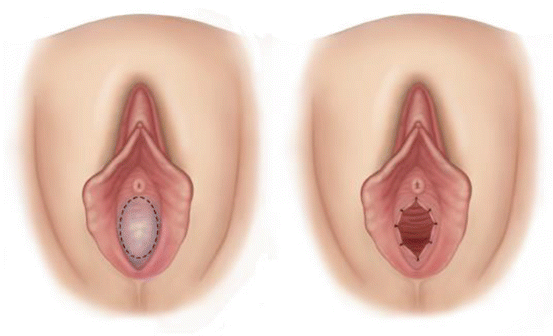
Fig. 3.1
Congenital anomalies of the hymen. (a) Normal hymen; (b) incomplete perforated hymen; (c) microperforate hymen; (d) cribriform hymen; (e) septate hymen
3.1.1 Diagnostic Criteria
3.1.1.1 Imperforate Hymen
Imperforate hymen is also known as nonporous hymen. The incidence of imperforate hymen is about 0.015 %. Imperforate hymen mainly obstructs the discharge of vaginal secretions. It may be asymptomatic at childhood due to minimal vaginal secretions, but at puberty, as both the vaginal and cervical secretions gradually increase, they accumulate in the vagina and lead to a sense of heaviness in the lower abdomen. After menarche, as the menstrual blood cannot drain out, it accumulates in the vagina forming a vaginal hematoma after several menstruations. Subsequently, it may lead to uterine and tubal hematomas, and eventually retrograde flow will enter into the pelvic cavity forming pelvic hematomas. Clinical symptoms are obvious with a cyclical lower abdominal pain, which progressively increases in intensity.
On gynecological examination, a bulging hymen can be seen, with a purple blue surface; on rectal examination, a vaginal mass bulging into the rectum is felt as a palpable pelvic mass. With a finger pressing on the vaginal mass, the bulging hymen can be made more obvious. Ultrasound scan may show accumulated fluid in the vagina and even in the uterine cavity.
3.1.1.2 Microperforate Hymen
It is a rare anomaly. There is a film covering the vaginal opening with just a pin hole opening as first described by Capraro in 1968 [1]. Since the pin hole is often difficult to detect, it is often misdiagnosed as an imperforate hymen. According to the sizes of the pin hole, the presenting symptoms will be different. Some patients have periodic menstruations with moderate amount of menstrual blood outflow, but a lot of blood still accumulates in the vagina; sometimes the periods are irregular; some patients presented with delayed menarche, with their chief complaints that include periodic abdominal pain and painful pelvic mass due to hematoma formation [2]. Microperforate or imperforate hymen can occur as an isolated anomaly, but it can also be associated with other reproductive tract anomalies, such as bicornuate uterus, vestibular anomalies, and anal atresia [3].
Similar to imperforate hymen, microperforate hymen can cause reflux of vaginal secretions and blood into the peritoneal cavity forming a pelvic mass. Unlike imperforate hymen, it will also cause recurrent urinary tract infections and sometimes a pelvic abscess. It is because the pinhole opening can have communication to the outside, through which bacteria will enter and multiply in the accumulated effusion or blood in the vagina or the pelvis leading a pelvic abscess. Bacteria can also migrate out of the vaginal opening, enter into the urethra, and cause recurrent urinary tract infections. On the contrary, patients with imperforate hymen do not have these presentations because their vaginas do not communicate with the outside [4]. The microperforate hymen is mainly diagnosed by examination under general anesthesia, and nowadays, fiber-optic hysteroscopy can be useful in its diagnosis together with simultaneous examination of any anomalies in the vagina and cervix [5].
3.1.2 Indication and Timing of Surgery
Surgery may be performed at any age. The ideal times are at the postneonatal period, puberty, or before menarche. As the development of hematocolpos can cause blood accumulation in the vagina, uterus, and fallopian tubes, followed by secondary endometriosis or pelvic infection, therefore, once hymen anomalies is diagnosed, surgery should be performed as soon as possible.
3.1.3 Surgical Contraindications
Vaginal atresia or the congenital absence of a vagina and other congenital anomalies should be ruled out before performing the surgery to incise the hymen. Surgery must be after a proper diagnosis of other anomalies (Fig. 3.2).


Fig. 3.2
Surgical incision of hymen
3.1.4 Preparative Preparation
Preoperative preparation is similar to other vulval surgery with routine cleansing and antiseptic preparation of the vulva.
3.1.5 Anesthesia and Positioning
The patient is placed in a lithotomy position. Local infiltration with anesthetic or intravenous anesthesia or general anesthesia can be used.
3.1.6 Surgical Procedure
Many literatures recommend the same surgical treatment for both microperforate and imperforate hymen. This includes surgical incision and removal of the accumulated blood in the vagina. After the surgery, patients will have significantly improved pregnancy rate and quality of life.
3.1.6.1 Incision
A metal urinary catheter should be used if possible to guide its position to avoid bladder injury. The surgeon should wear double gloves and insert the index finger of the left hand into the anus pressing towards the vagina for guidance, so as to avoid injury to the rectum. The incision should be at the most prominent part of the bulging hymen, though it will depend on the personal choice. The incision can be a “cross” incision, a vertical incision or a puncture incision at the center. Some surgeons think that it should begin with a small incision (especially for a thickened hymen), so as to reduce the rate of blood flow and hence to prevent vasovagal reaction due to the sudden decompression of the vaginal hematoma. Then, the outer glove of the left hand is removed to examine if the vaginal opening can accommodate one finger. Attention should be paid to avoid damaging the urethra and rectum during surgery. Finally, the vagina and cervix should be examined for other associated anomalies.
3.1.6.2 Evacuation of Hematoma
After opening the imperforate hymen, the retained menstrual blood will drain off. Any blood should be mopped up with gauze, and the cervix can now be visualized and examined. In the presence of a cervical adhesion or stenosis, a small uterine dilator is used to discharge any intrauterine blood retention. Tubal blood collection will gradually drain out following the operation. The abdomen should not be pressed or kneaded to avoid rupture of a hematoma or to force more retained blood flowing into the peritoneal cavity.
3.1.6.3 Suturing Edges of the Incisions
Any redundant hymen tissue should be cut off from the incision wounds. Absorbable sutures are used to suture the edges, while a very thin hymen wound without bleeding may not require suturing. In the literatures, absorbable sutures No. 3 “0” or No. 4 “0” are recommended to suture the wound edges or to cauterize wound edges with diathermy, so as to avoid wound adhesion with subsequent stenosis.
3.1.7 Postoperative Management
1.
Rest in a semisupine position is recommended, but patient is encouraged to sit up or to get out of bed to facilitate the drainage of any residual retained blood.
2.
It is important to maintain good vulval hygiene. Sitz bath or vaginal lavage is necessary for one week to avoid ascending infection. Prophylactic antibiotic is not often used.
3.
Patient can be discharged home on postoperative day 2 if she is fit for discharge.
4.
After 6–8 weeks, the uterus and the fallopian tubes will recover completely to their normal conditions. Any persistent enlargement of the fallopian tubes or persistent symptoms of peritoneal irritation should require further investigations.
3.2 Virilization of the External Genitalia
(3)
Department of Obstetrics and Gynecology, Peking Union Medical College Hospital, No. 1 Shuaifuyuan, Beijing, 100730, P. R. China
3.2.1 Perineal Incision and Repair
3.2.1.1 Diagnostic Criteria
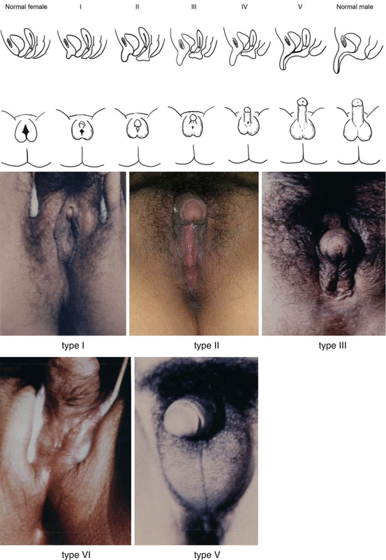
Female patients with a normal vagina but with a high perineal body or fused labia minora would have their vaginal opening completely or partially obliterated, resulting in poor menstrual blood flow or poor sex life. This vulval anomaly is mainly due to abnormal androgenic influence. Clinically it can be manifested as ambiguous external genitalia, labial fusion, and common vaginal and urethral opening. Prader classified external genital anomalies into the five types according to the varying degrees of virilization of the external genitalia: As illustrated by the following photos (Fig. 3.3), these types are described as:

Type I: Larger clitoris, with normal vagina and urethral openings.
Type II: Larger clitoris, with a funnel-shaped vaginal opening, but the openings for the vagina and urethra are still separated.
Type III: Significantly enlarged clitoris, both the vagina and urethra open from a common urogenital sinus.
Type IV: Significantly enlarged clitoris like a penis, the base of the penis is at the urogenital sinus, similar to hypospadias with fusion of the reproductive uplifted part.
Type V: Clitoris looks like a penis with the urethral opening at the penis tip. There is a complete fusion of the reproductive uplifted part; it is often mistaken as a male penis with cryptorchidism and hypospadias.

Fig. 3.3
Appearances of the external genitalia of normal female, 5 types of various degrees of virilisation and normal male
3.2.1.2 Operative Indications
Diagnosis can be easily made and accurately confirmed by examination. They are patients with a normal vagina and high vulval body or fusion of labia minora.
3.2.1.3 Timing of Surgery
1.
After menarche, patient presented with poor menstrual flow.
2.
Patient with difficult or impossible sexual intercourse.
3.
Patient for clitoral surgery.
3.2.1.4 Preoperative Preparation
Routine vulval skin cleaning and antiseptic preparation are performed.
3.2.1.5 Anesthesia
Local anesthesia is used for the labial incision and repair. If there is also a simultaneous corrective surgery for the clitoris or for vaginoplasty, general anesthesia or epidural anesthesia is recommended for these procedures.
Stay updated, free articles. Join our Telegram channel

Full access? Get Clinical Tree


