Introduction
Historically metatarsus adductus (MTA) was first described in Germany in the mid 19th century and only acknowledged in the English literature for the first time in 1921 (Berg 1986, Kite 1967). At times synonymous with metatarsus varus, MTA has long been a controversial entity in terms of treatment. In part, this confusion has been due to inconsistent identification of sub-types and thus a lack of helpful classification. Different types of MTA need to be identified as these need to be managed, and respond differently to treatment (Dietz 1994, Ganley 1984, Gore & Spencer 2004, Ponseti & Becker 1966, Sass & Hassan 2003). Fortunately the difficult types of MTA are in the minority and the clinical course is generally one of natural improvement (Widhe 1997). However, the less numerous, recalcitrant sub-types of MTA need to be carefully screened and managed to avert ongoing problems as the child grows (Gore & Spencer 2004; Sass & Hassan 2003; Tachdjian 1985, 1997).
Aetiology
Metatarsus adductus is defined as a transverse plane deformity at Lisfranc’s (the tarso-metatarso) joint in which the metatarsals and phalanges adduct from the rear foot, giving rise to a convex lateral border (Gore & Spencer 2004; Shepherd 1995; Tachdjian 1985, 1997; Tax 1980; Thomson 1993; Valmassy 1996).
The most cited causative factor is abnormal intrauterine position and resulting compression from the uterine wall against the developing foot (Valmassy 1996). Limited histological research from the second trimester has indicated abnormality of the medial cuneiform (Morcuende & Ponseti 1996). Other associations include (alone or in combination):
• tight abductor hallucis
• malpositioned tibialis anterior tendon at the base of the first ray
• tibialis anterior contracture (with weak peroneals)
• abnormal insertion of tibialis posterior
• arrested foot development in the first trimester (Valmassy 1996).
Prevalence
Metatarsus adductus is estimated to occur in 1:1000 births, making it a common foot deformity (Dietz 1994, Gore & Spencer 2004, Sass & Hassan 2003).
Twenty years ago, Berg reported that 56% of children referred to a particular institution in the USA had a diagnosis of metatarsus adductus (Berg 1986).
MTA has been reportedly more frequent in females, showing a predisposition for the left foot (Sass & Hassan 2003), yet others state there is no gender predilection (Valmassy 1996).
Some 50% of cases are bilateral and there is a 5% chance of subsequent siblings of affected children also having MTA (Drennan 1992).
Diagnosis
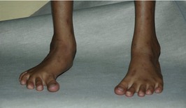
The diagnosis of MTA is clinical and includes a tell-tale ‘C’-shaped or ‘banana’ foot appearance. The small feet of neonates presenting with MTA morphology need to be carefully examined and classified, so that treatment is not withheld from the (fewer) types which do not otherwise resolve.
Key Concepts
Key Concepts
MTA can be usefully classified by three main attributes:
Classification
Classification by type
Metatarsus adductus is a broad generic term, used to describe a range of congenital foot deformities (Morcuende & Ponseti 1996). Clinically it is useful to sub-type MTA as follows:
1. Developmental metatarsus adductus (MTA type 1)
The developmental or postural type of MTA is positional in nature due to tightness of abductor hallucis, which may have been congenitally tight or as a result of intrauterine moulding. Fortunately this is the most common type of MTA and can usually be expected to spontaneously resolve. Manual stretching of the foot to a corrected posture at nappy changes may hasten this resolve. The metatarsals are in normal alignment and the adduction of the hallux (atavism) is only seen in dynamic simulated weight-bearing situations. The lateral border is typically straight.
2. True metatarsus adductus (MTA type 2)
This is the simplest of the structural types exhibiting transverse plane adduction of the forefoot on rear foot. All metatarsals are adducted, with the first being most adducted and the fifth being least adducted. The adduction is isolated at the Lisfranc joint (tarso-metatarso) and results in the ‘banana’ or ‘C’-shaped foot. The midfoot and rear foot are relatively normal (Drennan 1992).
3. Metatarsus (adducto) varus (MTA type 3)
This foot shows transverse plane adduction and also frontal plane inversion of the forefoot on rear foot. It involves all metatarsals being adducted at the Lisfranc joint and inverted distally. The calcaneus is generally everted, but can be straight. This type of MTA usually becomes a more entrenched deformity with time, so must be both recognized and treated effectively (Bleck 1971, Valmassy 1996).
4. Skew foot (MTA type 4)
This more complicated and – prognosis-wise – worse foot has transverse plane adduction and also frontal plane inversion of the forefoot on rear foot. Simultaneously, the rear foot is everted at both subtalar and midtarsal joints. This is a complex foot type, which in an older child or adult exhibits lateral midtarsal shift on X-ray. This type of MTA is usually acquired as either a compensation for uncorrected metatarsus varus (type 3) or severe true metatarsus adductus (type 2), but may also be induced by inappropriate treatment, for example forcibly abducting the forefoot without stabilizing the rear foot (Fig. 9.1; Ganley 1984).
Most importantly, you must notice that none of these deformities has a sagittal plane component, which if present will identify it as talipes equinovarus or ‘clubfoot’. Metatarsus adductus has been loosely termed ‘a third of a club-foot’ but does not exhibit ankle equinus (Ganley 1984, Kite 1967).
Classification by flexibility and degree of deformity
1. Flexibility
This is assessed by reducing the adduction of the forefoot on a stable rear foot in the transverse plane:
• Flexible: the deformity will reduce with ease, it being possible to abduct the forefoot beyond the foot’s midline.
• Reduced flexibility: the deformity will reduce no further than the foot’s midline with passive abductory force.
• Rigid: the deformity will not reduce and with abductory force applied, remains adducted, i.e. it does not reach the foot’s midline.
To determine overall flexibility it is also necessary to check the tension of abductor hallucis in both non-weight-bearing and also weight-bearing (simulated if baby is pre-walking). It is common with a tight abductor hallucis belly to see a flexible metatarsus adductus exhibit marked adduction of the hallux in weight-bearing and gait. This may be termed dynamic hallux varus or an atavistic first ray.
Clinical flexibility guides treatment. Structurally fixed cases will not reduce passively and may require surgical correction.
2. Degree of deformity
Bleck’s (1983) simple clinical scale records the severity of metatarsus adductus from the child’s footprint. The heel is bisected and a line extended through the forefoot (Fig. 9.2). Bleck’s scale can be adapted for clinical use as shown below. Clinically, the use of a ruler is useful for this method.
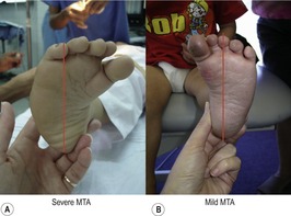
This method allows for simple non-invasive baseline assessment and monitoring of the MTA deformity and classification of the degree of deformity:
| i. | normal | heel bisection ends distally at | 2nd/3rd toes |
| ii. | mild | heel bisection ends distally at | 3rd toe |
| iii. | moderate | heel bisection ends distally at | 3rd/4th toes |
| iv. | severe | heel bisection ends distally at | 4th/5th toes laterally. |
Differential diagnosis
• Clubfoot (talipes equinovarus).
• Metatarsus primus varus.
• Cerebral palsy.
Typical clinical picture
Examination
Examination and diagnosis of metatarsus adductus is based on clinical appearance in the infant and young child. The prominence of the styloid process of the fifth metatarsal is a hallmark sign for adduction of all five metatarsals versus the so-called atavistic first ray where the lateral border of the foot is straight but the medial concave/adducted. Skin creasing on the medial side is usually associated with a less flexible MTA deformity which is more likely to require treatment. There should be no reduction of ankle joint dorsiflexion with MTA. If ankle range is reduced, you need to suspect talipes equinovarus or clubfoot.
Key Concepts
Key Concepts
Atavistic first ray refers to a primitive foot type where the first ray acts more like a thumb with grasping ability. Useful for hanging from trees originally, this can clinically contribute to an increased or ‘dynamic’ MTA with weight-bearing where the first ray adducts and can medially rotate the lower leg and contribute to an intoeing gait. Shoes with a straight medial border seem to reduce the action of abductor hallucis and in turn reduce the hallux adduction grasp and intoeing.
Radiological examination is useful in later stages to classify the extent of deformity based on specific joint angle deviations and values (Table 9.1).
| Angle | Normal values | |
|---|---|---|
| Metatarsus adductus angle (AP) | Angle formed between longitudinal bisection of second metatarsal and the midtarsal divide The midfoot divide is difficult to identify prior to ossification | Birth: 22–25° Toddler:15–20° Adult: 10° |
| Talo-first metatarsal angle (AP) | Angle formed between talar bisection and first metatarsal bisection Useful as a basic evaluation of atavistic first metatarsal versus forefoot adductus | 0–20° |
| Talo-calcaneal angle (AP) | Angle formed between talar and calcaneal bisections Useful in discrimination from clubfoot < div class='tao-gold-member'>
Only gold members can continue reading. Log In or Register to continue
Stay updated, free articles. Join our Telegram channel
Full access? Get Clinical Tree


|