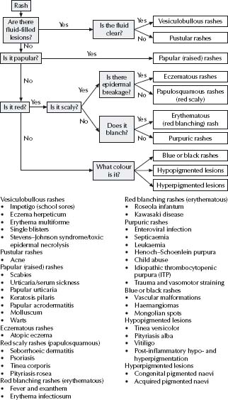
Vesiculobullous rashes
Vesicles are usually caused by infections (herpes simplex virus (HSV), varicella zoster virus (VZV), enterovirus, tinea, scabies or impetigo) or contact dermatitis. Also, consider drug reactions and erythema multiforme. Larger blisters may be from staphylococcal infections, tinea, Stevens–Johnson syndrome, arthropod bites, contact dermatitis, burns or trauma.
Impetigo (school sores)
Cause
Staphylococcus aureus or Streptococcus pyogenes (impetigo but not bullous lesions), or both.
Clinical features
Impetigo presents as areas of ooze and honey-coloured crusts on the face, trunk or limbs. Occasionally, the primary lesions are bullous. Lesions are rounded and well demarcated and are most often grouped and asymmetrical but may be solitary and widespread. Their onset and spread may be rapid or occur over days. In more chronic cases, there may be central healing with peripheral spread to give annular lesions.
Management
- Bathe off crusts.
- Apply topical mupirocin 2% ointment 8 hourly if localised, or cephalexin 25 mg/kg (maximum 500 mg) orally, 12 hourly if severe or extensive.
- Isolate the child from other children or from sick adults unless all lesions are covered or treated.
- Treat any underlying condition such as scabies (a common cause of widespread impetigo) or eczema.
Staphylococcal scalded skin syndrome
Clinical features
- Usually seen in younger children.
- Mediated by an epidermolytic toxin released from an often insignificant staphylococcal focus (e.g. eyes, nose or skin).
- Fever and tender erythematous skin are early features.
- Exudation and crusting develops, especially around the mouth.
- Wrinkling, fl accid bullae and exfoliation of the skin are seen – Nikolsky sign (‘normal’ skin separates if rubbed).
- Blisters are very superficial and heal without scarring.
Management
- Flucloxacillin 50 mg/kg (max 2 g) i.v. 6 hourly if there is evidence of sepsis or systemic involvement.
- Look for a focus of infection. Drain foci of pus if present.
- Monitor temperature, fluids and electrolytes if large areas are involved. Increased fluids will aid renal excretion of toxin.
- Handle skin carefully and use an emollient ointment.
Erythema multiforme
Clinical features
This is a specific hypersensitivity syndrome that occurs at any age. Lesions are usually symmetric and appear most commonly on the hands, feet and often the face. They can be found anywhere, including mucous membranes.
Typical target lesions have an inner zone of epidermal injury (purpura, necrosis or vesicle), an outer zone of erythema and sometimes a middle zone of pale oedema. They are not migratory. Most cases are caused by herpes simplex, some by other infections. Drugs are an uncommon cause.
Management
- Fluid maintenance.
- Apply emollient ointment to the lips, if needed.
- If the condition is recurrent, it is highly likely to be related to HSV. Prophylactic aciclovir should be considered if recurrences are frequent and affecting the quality of life.
Stevens–Johnson syndrome/toxic epidermal necrolysis
Clinical features
Stevens–Johnson syndrome and toxic epidermal necrolysis are believed by many to be variants of the same condition.
- They are characterised by widespread blisters on an erythematous or purpuric macular background, often with extensive mucous membrane haemorrhagic crusting.
- There may be tender erythematous areas with a positive Nikolsky sign (‘normal’ skin separates if rubbed).
- Conjunctivitis, corneal ulceration and blindness can occur. Some degree of permanent scarring around the conjunctivae is common even if eye symptoms are not severe.
- Anogenital lesions can lead to urinary retention.
- Fever, myalgia, arthralgia and other organ involvement can occur.
- Drugs are the most common cause, occasionally Mycoplasma.
Management
- Cease any drug that may be the cause.
- Fluid maintenance. Monitor temperature, fluids and electrolytes.
- Apply emollient ointment to the skin, lips and anogenital areas – this may be required many times a day. Careful attention to emollients and/or dressing of the glans and under-surface of the foreskin may prevent secondary scarring, adhesions and phimosis.
- A regular eye examination with a specialist review for topical steroid drops if any eye involvement is noted.
- Good pain management is essential. Occasional or regular brief inhalational general anaesthetics may be necessary to facilitate dressings, eye care, etc., which must not be compromised.
- I.v. gammaglobulin is seen by many as standard therapy for cases threatening to become severe.
- Cyclosporin (5–6 mg/kg per day for a few days, then taper to 3–5 mg/kg per day for 2–3 weeks) commenced at diagnosis may prevent worsening. Immunosuppression is controversial (beware sepsis).
Note: Stevens–Johnson syndrome is not severe erythema multiforme (EM). They are distinct conditions with different aetiologies. Permanent sequelae are rarely seen in severe EM and concurrent drug use is unlikely to be the cause. Skin lesion morphology is the best discriminating factor. Classic target lesions are not seen in Stevens–Johnson syndrome. Mucous membrane involvement can be seen in both conditions but is usually localised in EM, often confl uent in Stevens–Johnson syndrome.
Eczema herpeticum
Clinical features
HSV infection in children with eczema is common, but many cases are misdiagnosed as either an exacerbation of the eczema or bacterial infection. Grouped vesicles may be prominent, but more often vesicles are rudimentary or absent and the infection presents as a group of shallow 2–4 mm ulcers on an infl amed base. The infected area may not be painful or itchy and does not respond to standard eczema therapy. If untreated, resolution usually occurs in 1–4 weeks, but dissemination may occur. Recurrences may occur at different sites.
Management
- Collect epithelial cells from the base and roof of the vesicles for herpes immunofluorescence and culture.
- Local disease in an otherwise well child requires regular observation but does not need antiviral therapy.
- A child with fever or multiple sites of cutaneous herpes infection may need admission to hospital and treatment with i.v. aciclovir 20 mg/kg per dose (2–12 weeks), 250 mg/m2 per dose (12 weeks–12 years), 5 mg/kg per dose (>12 years) 8 hourly i.v. over 1 h.
- Milder cases demonstrating progression or facial involvement can be managed with oral aciclovir.
- Eye involvement should be managed with topical or systemic aciclovir, or both, and urgent review by an ophthalmologist.
- The underlying eczema can be treated with moisturisers, topical steroids and wet dressings.
Single blisters
When a child presents with a single blister as an isolated finding, consider impetigo, tinea, mastocytoma, insect bite, cigarette burn, friction or spider bite. The latter can grow over days to become a non-tender blister with a diameter of many centimetres.
Consider acne, folliculitis, scabies, perioral dermatitis, acute generalised exanthematous pustulosis and rarely psoriasis.
Acne
Clinical features
Acne mainly affects the forehead and face but can involve other sebaceous gland areas (neck, shoulders and upper trunk). Early lesions include blackheads, whiteheads and papules. In more severe cases there may be pustules or inflammatory cysts that can lead to permanent scarring. Undertreated acne is a cause of significant morbidity in adolescents and may be a factor in teenage suicide. Consider underlying endocrine disorders if acne begins before puberty.
Management
- Acne is treatable. No person with acne should just be told it is an inevitable part of adolescence. Effective acne therapies are now available and should be used to control the disease.
- For mild disease, use topical benzoyl peroxide 2.5–5%. Other topical agents include antibiotics (clindamycin, erythromycin or tetracycline), isotretinoin (not in pregnancy) or azelaic acid. These can be used singly or in combination. Improvement occurs over 1–2 months,
- not within days. All of these topical agents have side effects with which practitioners must be familiar.
- Treatment of moderate acne often involves the addition of oral antibiotic therapy (e.g. tetracycline 500 mg twice daily, erythromycin 500 mg twice daily, doxycycline 50–200 mg per day) for 3–6 months.
- Oral hormone therapy can help female patients.
If antibiotics and topical treatment have not resulted in considerable improvement in 3 months, oral isotretinoin (Roaccutane®) is indicated. Provided pregnancy is avoided, this is safe and highly effective. Isotretinoin is also indicated if there is scarring, cyst formation, or significant depression. Treatment of severe acne with isotretinoin may decrease the risk of suicide. Assessment and treatment of any depression is also required.
If the child is itchy, consider scabies, urticaria, serum sickness, papular urticaria or molluscum. If not, consider urticaria, molluscum, warts, melanocytic naevi, keratosis pilaris and papular acrodermatitis. For vascular swellings, consider pyogenic granuloma.
Scabies
Clinical features
An intensely itchy papular eruption develops 2–6 weeks after first exposure to the Sarcoptes scabiei mite or 1–4 days after subsequent reinfestation. The characteristic lesion is the burrow that is several millimetres in length. Burrows are best seen on the hands, especially between the fingers, and on the feet. Early burrows may be vesicular. A clue to the diagnosis of scabies is the distribution of papules and pruritus. Involvement of the palms, soles, axilla, umbilicus, groin and genitalia is common and the head is usually spared. Excoriations and secondary impetigo may be present. There is currently a worldwide pandemic of this contagious disease affecting both adults and children.
Management
- Treatment is expensive and upsetting. If diagnosis is unclear, confirm by scraping to find a mite, or refer before treating.
- Use permethrin 5% cream. An alternative for pregnant women or neonates is sulfur 2% in yellow soft paraffin.
Note: The following are not recommended: lindane 1% (contra-indicated in infants or women who are pregnant or breast-feeding) or benzyl benzoate 25% (too irritating for children and ineffective if diluted).
- Treat all family members and any other people who have close skin contact with the affected individuals.
- Apply to dry skin (not after a bath) from the neck down to all skin surfaces. For infants, apply to the scalp as well (not face). Use mittens if necessary to prevent finger sucking.
- Leave the cream on for at least 8 h.
- Wash the cream off. Wash clothing, pyjamas and bed linen at this time.
- The itch takes a week or two to settle and can be treated with potent topical steroids.
- Reinfestation is common. The family should notify all social contacts (e.g. crèche, school or close friends) to ensure that all those infected receive treatment.
Urticaria/serum sickness
See also chapter 19, Allergy and immunology.
Clinical features
Urticaria is characterised by the rapid appearance and disappearance of multiple raised red wheals on any part of the body. Individual lesions are often itchy and clear within 1 day. There may be central clearing to give ring lesions (these are not the so-called target lesions of EM that persist for several days). The child is usually well. Urticarial episodes usually resolve over days or weeks and rarely last longer than 6 months. In most cases of short duration, the trigger is either a transient viral infection, allergic reaction or cannot be determined.
Some children develop fever and arthralgias in association with urticarial lesions that are more fixed and may bruise or be tender (serum sickness). In Australia, serum sickness is usually idiopathic or following a course of cefaclor.
Management
- Urticaria may be the first sign of anaphylaxis. If there is associated angio-oedema (prominent subcutaneous swelling) or wheeze, continued observation and appropriate treatment is required (see chapter 1, Medical emergencies).
- Investigation is usually not required.
- Ask about medications; new foods and environmental allergens.
- Treat the itch with oral antihistamine (see chapter 19, Allergy and immunology, p. 228). Oral prednisolone (1 mg/kg per day, max. 50 mg) for 2–5 days is beneficial in serum sickness and is warranted in urticaria when pruritus is severe.
- Urticaria can become chronic, and in the vast majority of cases no underlying ongoing trigger is found. Consider mast cell degranulating drugs, foods, animals, parasitic infections, heat, cold and physical pressure. Consider investigating with a throat swab (for streptococcal carriage), full blood examination (for eosinophilia and anaemia), antinuclear antibodies, urine culture for bacteriuria, nocturnal check for threadworms and a possible challenge with any suspected agent. Adding cimetidine (10 mg/kg (max 200 mg) p.o. 6 hourly) to the antihistamine may help.
- If individual lesions last >2 days or are tender or purpuric, consider investigation for cutaneous vasculitis.
Papular urticaria
Clinical features
This is a clinical hypersensitivity to insect bites. New bites appear as groups of small red papules, usually in warmer weather. Older bites appear as 1–5 mm papules, sometimes with surface scale or crust, or with surrounding urticaria. Vesicles or pustules may form. Individual lesions may resolve in a week or last for months and may repeatedly fl are up after fresh bites elsewhere. The itch is often intense and secondary ulceration or infection is common.
Management
- Prevent bites (e.g. adequate clothing, modifying behaviour that leads to exposure, occasional repellent and the treatment of pets and house for fleas if necessary).
- Treat the itch with an agent such as aluminium sulfate 20% (Stingose), liquor picis carbonis 2% in calamine lotion, potent steroid ointment or antihistamines (see chapter 19, Allergy and immunology, p. 228). Protective dressings (e.g. Duoderm) can speed the healing of lesions.
- Treat secondary infection with topical mupirocin ointment 2% or oral antibiotics.
Keratosis pilaris
Clinical features
This is a rough, somewhat spiky papular rash on the upper outer arms, thighs, cheeks, or all three areas, with variable erythema. It is common at all ages.
Management
- Reassure the patient that this is rarely a problem. Soap avoidance and moisturisers can improve the feel. Steroids don’t help.
- Older children may get some benefit from topical keratolytics (e.g. Dermadrate, Calmurid).
- Older children with troublesome facial redness can be treated with vascular laser (V beam).
Papular acrodermatitis
Clinical features
This is characterised by the acute onset of monomorphic red or skin-coloured papules mainly on the arms, legs and face. It is usually asymptomatic. It can be caused by coxsackie virus, echovirus, mycoplasma, EBV, adenovirus and others.
Management
Reassure and advise that clearing can take several weeks.
Molluscum
Clinical features
Uncomplicated molluscum lesions are easily recognised as firm, pearly, dome-shaped papules with central umbilication; however, presentation to a doctor is often prompted by the development of eczema in surrounding skin. In such cases, recognition can be difficult as eczematous changes can obliterate the primary lesions. A careful history of the initial lesions is usually diagnostic.
Management
Education – molluscum is caused by a virus and is very common. A child may develop a few, or a great many lesions and individual lesions may last for months. Complete resolution will not happen until an immune response develops, which may take from 3 months to 3 years.
Children with molluscum should not share towels but should not be restricted in their activities.
The treatment depends on the age of the child, the location of the lesions and any secondary changes. Things to note include:
- Treatment of the surrounding eczema may be all that is required.
- Uncomplicated lesions not causing problems and not spreading can be left alone.
- Isolated or troublesome lesions (e.g. on the face) can be physically treated. One method is gentle cryotherapy.
- Rarely, children warrant curettage under topical anaesthesia. This is well tolerated and usually curative but can potentially scar. Alternatively, the stimulation of an immune response can be attempted with cantharidin, aluminium acetate solution (Burow’s solution 1:30) for large areas, or benzoyl peroxide 5% daily to small areas and covered with the adhesive part of a dressing.
- Inflamed lesions do not require antibiotic treatment but if true cellulitis or abscess formation occurs, treat with antibiotics and/or drainage.
Warts
Many serotypes of the papilloma virus can cause warts. Different serotypes have a predilection for different areas of the skin. No treatment is necessary unless the warts are causing a problem to the child (e.g. social embarrassment, or pain from a plantar wart). Avoid painful procedures unless chosen by older children. Resistant warts on the limbs often respond to contact sensitisation (e.g. Diphenylcyclopropenone (DCP) 0.1% cream after sensitisation with 2% solution). Diphenylcyclopropenone use requires caution, supervision and possible dose adjustment, as there is a wide variation in individual responses.
- Ordinary warts: if tolerated by the child, paring every 2–3 days with a razor blade or nail file will remove the surface horn. Apply a proprietary keratolytic agent that contains salicylic or lactic acid, or both, each day or two as directed.
- Plantar warts: these can be painful and can appear fl at. Pare as for ordinary warts. Apply a proprietary keratolytic agent that contains salicylic or lactic acid, or both, each day or two. Alternatively, place a small pad of cotton wool soaked in 3% formalin in a saucer on the floor. Rest the wart-affected sole on the pad/saucer for 30 min each night. Cryotherapy and surgery are often ineffective and can lead to painful keloid scarring.
- Plane (flat) warts: these are smooth, fl at or slightly elevated, skin-coloured or pigmented lesions. They may occur in lines or coalesce to form plaque-like lesions. If treatment is needed for plane warts on the hands, apply a formalin solution as for ordinary warts. Lesions on the face are often subtle and may not need treatment. Treatment may cause complications such as pigmentary changes and requires considerable caution.
- Anogenital warts: these are soft, fleshy warts that occur at the mucocutaneous junctions, especially around the anus. They may be isolated flesh-coloured nodules or may coalesce into large cauliflower-like masses. Management options include awaiting resolution, topical podophyllotoxin, imiquimod, curettage and diathermy and carbon dioxide laser.
Note: the presence of genital warts in a young child is not an indication for mandatory reporting to government protective services. Genital warts in children should lead to consideration of sexual abuse, but transmission is usually by normal close parent–child contact.
Consider atopic eczema, allergic contact dermatitis, irritant contact dermatitis, photosensitivity eruptions, molluscum, tinea corporis and scabies.
Atopic eczema
See also chapter 19, Allergy and immunology.
Clinical features
Eczema usually begins in infancy. It commonly involves the face and often the trunk and limbs as well. In older children the rash may be widespread or may be localised to flexures. Erythema, weeping, excoriation and rarely vesicles may be seen in acute lesions. Chronic lesions may show scale and lichenification. In some children the lesions are more clearly defined, thickened discoid areas that may intermittently be itchy. There is usually a cyclical pattern of improvement and exacerbation. Weeping and yellow-crusted areas that do not respond to therapy may indicate secondary bacterial or herpetic infection.
Management
- Education: parents need to know the triggers and that treatments are effective in controlling the disease.
- Avoid irritants which may worsen eczema: soaps, bubble baths, prickly clothing, seams and labels on clothing, car seat covers, sand, carpets, overheating or contact with pets. Smooth cotton clothing is preferred.
- Keep the skin moist: use a moisturiser such as paraffin ointment (50:50 white soft paraffin/liquid paraffin) as often as several times a day if necessary.
- Treat inflammation: in mild or moderate cases, steroid creams can be used intermittently with good effect. Hydrocortisone 1% is usually adequate. If not, moderate potency (e.g. beta-methasone valerate 0.02%) or potent (e.g. mometasone 0.1% or methylprednisolone 0.1%) ointment can be used for exacerbations in areas other than the face or nappy area. Prolonged regular use of moderate-potency steroids to the skin of young children can cause atrophy and adrenal suppression. Oral steroids are rarely indicated in eczema. For chronic eczema on the limbs, zinc and tar combinations are alternatives to steroids.
- Control itch: advise parents to avoid saying ‘Stop scratching’ all the time, and to distract the child instead. Avoid overheating, particularly at night. Wet bandaging is very helpful if warranted. Antihistamines are often unhelpful but may be tried if the itch is not controlled by other measures (see chapter 19, Allergy and immunology). Note: Terfenadine (Teldane) and astemizole (Hismanal) should not be used because of occasional fatal interactions if erythromycin is also taken.
- Treat infection: take cultures and treat with simple wet dressings and oral antibiotics (e.g. erythromycin, cephalexin or fl ucloxacillin). Consider if herpes simplex is present (see p.271). For recurrent bacterial infection, use antiseptic wash or bath oil (e.g. triclosan).
- Diet: a normal diet is usually indicated. If a child has immediate urticarial reactions to a particular food, that food should be avoided. Environmental and food allergens may contribute to the exacerbation of symptoms in some patients. Allergen avoidance in these children may be of some benefit. In difficult cases, consider a more formal allergy assessment.
- Hospitalisation: if a child is missing school because of eczema, they should generally be in hospital for intensive treatment.
Red scaly rashes (papulosquamous)
Consider seborrhoeic dermatitis (infants), psoriasis, tinea corporis, pityriasis rosea, pityriasis versicolor and atopic eczema. Ichthyosis vulgaris is a common cause of generalised scale without itch or redness.
Seborrhoeic dermatitis
Clinical features
- This condition presents in the first months of life, partly due to the activity of commensal yeasts.
- Red or yellow/brown scaly areas will commonly affect the scalp and forehead. (The ‘seborrhoeic’ rash in infants affecting areas without sebaceous glands (e.g. axillae, napkin area) is probably best considered as a form of psoriasis.) The folds behind the ears and around the neck, axillae, groin and gluteal clefts are also affected.
- Resolution by the age of 1 year is usual.
Management
- Paraffin or olive oil applied to scalp to loosen scale.
- Imidazole creams with hydrocortisone 1% cream or with a mixture of salicylic acid (1%) and sulfur (1%) ointment, twice daily.
- Anti-yeast shampoos (e.g. selenium sulfide – Selsun) can be helpful. Use carefully to avoid irritation or toxicity.
Psoriasis
Clinical features
Psoriasis can occur at any age. Lesions begin as small red papules that develop into circular, sharply demarcated erythematous patches with prominent silvery scale. Common presentations include plaques on extensor surfaces, generalised guttate (small) lesions, red scaly scalp lesions or moist red anogenital rashes. Itch can be a variable feature. Nail changes are often seen in childhood.
Management
The treatment depends on the site and extent of disease and the age of the child. Adolescents are less tolerant of tar creams.
- Treat isolated skin plaques with either topical steroids (e.g. intermittent mometasone with clinical monitoring) or tar-based creams (e.g. liquor picis carbonis 3%, salicylic acid 2% in sorbolene cream). Generally avoid tars on the face, flexures and genitalia.
- Use hydrocortisone 1% ointment on the face and anogenital region. Topical steroids are not used for large areas in childhood psoriasis because of the possible development of rebound pustular disease.
- Thick scalp plaques can be softened overnight with a similar tar cream and removed with a tar shampoo.
- Topical calcipotriol can be used in conjunction with steroid creams.
- Widespread psoriasis may need treatment with one or more of dithranol, etretinate, methotrexate, cyclosporin or ultraviolet therapy, all of which are effective.
Tinea corporis
Clinical features
The typical lesion is a slow-growing erythematous ring with a clear or scaly centre; however, tinea corporis can present in a wide variety of ways, particularly if previously treated with steroid ointments. It can be pustular, vesicular or bullous, or spread to many sites within days. Tinea should be considered in any red scaly rash where the diagnosis is unclear.
Management
- If in doubt about the diagnosis, confirm by scraping the scale for microscopy and culture.
- Lesions are treated with terbinafine cream (twice daily for 1 week) or an imidazole cream (e.g. clotrimazole, miconazole or econazole 2–4 times daily, for 4 weeks).
- Oral griseofulvin (20 mg/kg per day in divided doses) is required for tinea capitis or for widespread lesions.
Pityriasis rosea
Clinical features
The condition is common between the ages of 1 and 10 years. Initially, a pink scaly patch appears, followed a few days later by many pink/red scaly oval macules mainly on the trunk. It is usually asymptomatic but can be itchy.
Management
Reassure the patient. The condition can persist for weeks.
Red blanching rashes (erythematous)
Macular erythematous lesions are most commonly caused by viral infections (e.g. coxsackie, echovirus, Epstein–Barr virus, adenovirus, parainfluenza, influenza, parvovirus B19, human herpes virus 6, rubella and measles) or drug reactions. Consider also septicaemia, scarlet fever, Kawasaki disease (see chapter 30, Infectious diseases) and Mycoplasma infection.
Fever and exanthem
The onset of fever and exanthem is usually due to a viral illness, often enterovirus. Some infections have specific clinical features that aid diagnosis; for example measles and erythema infectiosum. However, in most instances a diagnosis cannot be made with certainty. To manage such a child, consider:
- Is the child sick? Is the child lethargic, cold peripherally or young? Consider meningococcal disease, other bacterial sepsis and Kawasaki disease. Investigate and treat.
- Are they taking any medication? Consider ceasing medication.
- Are there other people at risk? If relatives are immunosuppressed or pregnant, consider serology, stool viral culture and advising the at-risk person to consult their doctor.
- Is the rash papular? Consider papular acrodermatitis.
If the answer to all the above is ‘no’, reassurance and review is probably appropriate.
Erythema infectiosum and Kawasaki disease
See chapter 30, Infectious diseases.
Roseola infantum
This condition is seen every day in paediatric emergency departments. Typically, an infant has had a high fever for 2–4 days and has often been put on antibiotics. The fever then goes but a widespread erythematous rash appears. The family need reassurance that the rash is not a drug reaction. See chapter 30, Infectious diseases.
Consider viral infections, meningococcal sepsis, platelet disorders, vasculitis, drug reactions and trauma.
Septicaemia
Suspect septicaemia (usually meningococcal) in a child with recent onset of fever and lethargy. Skin lesions may be erythematous macules progressing to extensive purple purpura. Even if there is doubt, take blood cultures, give antibiotics and arrange admission (see also chapter 1, Medical emergencies).
Enteroviral infection
Scattered petechiae are common in children who have fever from enteroviral infections. These children are usually well. If in doubt, or if the child appears unwell, investigate (full blood examination, blood cultures) and consider treatment for septicaemia. See Approach to the febrile child, p. 381.
Leukaemia
Suspect leukaemia in a child with generalised petechiae or purpura in the absence of trauma. Look for tiredness or pallor. Obtain an urgent full blood examination (see chapter 29, Haematologic conditions and oncology).
Henoch–Schönlein purpura
See detailed summary including Investigations and Management in chapter 37, Rheumatologic conditions.
Non-itchy, painless macules, papules or urticarial lesions with purpuric centres occur in a symmetrical distribution mainly on the buttocks and ankles, occasionally on the legs, arms and elsewhere. There may be associated abdominal pain, arthralgia, arthritis or haematuria. Renal involvement leading to chronic renal failure is rare, but can occur irrespective of the severity of the rash and other symptoms and may be delayed until weeks or months after the onset of the illness.
Idiopathic thrombocytopenic purpura
See also chapter 29, Haematologic conditions and oncology.
Bruises, petechiae or purpuric lesions appear over a period of days or weeks, mainly in sites of frequent mild trauma. The child is otherwise well. Full blood examination will show a low platelet count.
Child abuse
See chapter 17, Child abuse.
Twisting, compression, pinching and hitting can all cause petechial or purpuric lesions. Look for bruises of bizarre shapes and different ages, evidence of bony fractures and an abnormal affect.
Trauma and vasomotor straining
In some ethnic groups it is common to treat a febrile or unwell child by rubbing or suctioning the skin with a variety of implements. This produces bizarre circular and linear patterns of petechiae that can alarm the unwary.
Petechiae can appear around the head and neck in normal children after coughing or vomiting. Restraining a small child for a procedure such as a lumbar puncture or venepuncture can also lead to the development of petechiae on the upper body.
Consider vascular malformations, haemangiomas, Mongolian spots, blue naeviand melanoma.
Vascular malformations
- These can be blue, red, purple or skin coloured. They are developmental defects and do not resolve.
- Such malformations can involve any mix of capillaries (e.g. portwine stain), veins, arteries (e.g. arteriovenous malformation) and lymphatics (e.g. cystic hygroma).
- Extensive malformations can be associated with pain, soft tissue or bony hypertrophy, bone erosion, haemorrhage, infection and platelet trapping.
- Management requires a multidisciplinary approach using expertise from surgical, paediatric, dermatological and radiological fields.
Haemangiomas
Clinical features
Superficial haemangiomas begin as macular erythematous lesions in the first weeks of life and become soft, partly compressible, sharply defined, red or purple swellings that can occur anywhere on the body. Deeper houraemangiomas may appear as blue or skin-coloured swellings. Most haemangiomas are not present at birth; they grow for several months and resolve fully over several years.
Management
Parents need reassurance about the inherently benign nature of these lesions. Most haemangiomas are best left alone and allowed to involute spontaneously. In some sites, however, haemangiomas can rapidly lead to problems such as ulceration, blindness, destruction of cartilage, respiratory obstruction or death.
Urgent assessment by an experienced clinician is needed if any developing haemangioma:
- Is ulcerating and potentially disfiguring.
- Is on the eyelid or adjacent to the globe of the eye.
- Deforms structures such as the lip, ear cartilage or nasal cartilage.
- Begins as an extensive macule that grows thicker.
- Is associated with stridor, thrombocytopenia or multiple lesions.
Corticosteroids are usually used, occasionally with vascular laser, surgery or interferon.
In hypopigmented lesions, look for a fine scale. If it is scaly, consider pityriasis versicolor or pityriasis alba. If it is not scaly, consider pityriasis versicolor, post-inflammatory loss of pigment, halo naevi or vitiligo.
Pityriasis versicolor
- This is common in adolescents and is caused by an increased activity of commensal yeasts.
- Multiple oval macules, usually covered with fine scale, appear on the trunk or upper arms. The lesions may appear paler or darker than the surrounding skin.
- Treatment with anti-yeast shampoos is effective. For example, apply selenium sulfide 2% (Selsun shampoo). Leave on for 5–10 min, rinse and treat weekly for 4 weeks and then monthly. The pigmentation takes weeks to resolve and relapses are common without ongoing maintenance.
Pityriasis alba
This condition is common in prepubertal children and represents post-inflammatory hypopigmentation secondary to mild eczema. Single or multiple, poorly demarcated hypopigmented 1–2 cm macules are seen on the face or upper body. Lesions are not itchy but often have a fine scale. Reassure and treat with hydrocortisone 1% to active lesions and educate regarding skin care for eczema. Resolution of the discolouration takes weeks.
Vitiligo
This condition is characterised by sharply demarcated, often symmetrical areas of complete pigment loss. Eventual repigmentation in childhood vitiligo is common and is helped by topical steroids. In troublesome cases refer to a specialist for advice regarding treatment (e.g. corrective cosmetics or psoralen therapy).
Post-inflammatory pigmentation changes
This condition occurs particularly in dark-skinned people. Many inflammatory skin disorders may heal leaving diffuse, hypo- or hyperpigmented macules that can persist for months or years. No treatment is satisfactory.
If they are flat, consider junctional melanocytic naevi, café-au-lait spots, naevus spilus, pityriasis versicolor and post-inflammatory hyperpigmentation. If raised, consider compound melanocytic naevi, Spitz naevi and warts.
Congenital pigmented naevi
Congenital melanocytic naevi that cover large areas or are likely to cause significant cosmetic concern need very early assessment by a skin specialist and plastic surgeon, preferably in the first week of life, for diagnosis, surgery, laser treatment and/or long-term follow up. Small congenital melanocytic naevi have no increased risk for the development of melanoma over other moles.
Acquired pigmented naevi
During childhood, most children develop multiple pigmented lesions, which may be freckles, lentigines, naevus spilus, acquired melanocytic naevior very rarely, melanoma.
Immune-suppressed children and those who have had chemotherapy are at greater risk of skin malignancy.
Most anogenital rashes seen in infants who wear nappies are primarily caused by reaction with urine or faeces (irritant napkin dermatitis) or by seborrhoeic dermatitis. Soaps, detergents and secondary yeast infection may contribute. In older children, threadworms (Enterobius vermicularis) are a common cause of an itchy anogenital rash. Look for the worms at night and treat with mebendazole 50 mg (<10 kg), 100 mg (>10 kg) (not in pregnancy or <6 months) or pyrantel 10 mg/kg (max 500 mg) once oral. A repeat dose 2 weeks later helps reduce the high rate of reinfestation.
Consider also less common causes such as malabsorption syndromes (diarrhoea, erosive dermatitis and failure to thrive), zinc deficiency (a sharply defined anogenital rash with associated perioral, hand and foot ‘eczema’), Langerhans’ cell histiocytosis, psoriasis and Crohn’s disease.
Irritant napkin dermatitis
Clinical features
This is the most common cause of napkin dermatitis in infants and typically presents as confluent erythema that typically, but not always, spares the groin folds. Variant presentations include multiple erosions and ulcers, scaly or glazed erythema and satellite lesions at the periphery. Satellite lesions are suggestive of Candida infection.
Management
- Keep the area clean and dry. Leave the nappy off whenever possible.
- Gel-based disposable nappies or a non-wettable under-napkin can be helpful. Cloth nappies should be thoroughly washed and rinsed.
- Use topical zinc cream or paste for mild eruptions.
- Add hydrocortisone 1% cream if inflamed. Do not use stronger steroids.
- Consider mupirocin 2% cream if not settling. Antifungal therapy is often not needed, even if Candida is present.
Candida napkin dermatitis
This occurs secondary to irritant napkin dermatitis and antibiotic use. Swab to confirm and treat the underlying cause as above and use topical imidazole cream.
Perianal streptococcal dermatitis
Streptococcus pyogenes infection.
Clinical features
- A localised, well-demarcated erythema that covers a circular area of 1–2 cm radius around the anus.
- Tenderness and painful defecation are typical.
- If not treated, it may persist for months.
- May have fissures and constipation.
Management
- Take perianal and throat cultures to confirm the presence of Streptococcus pyogenes.
- Apply paraffin ointment three times daily to the perianal area for symptomatic relief. Treat with oral antibiotics (phenoxymethylpenicillin 15 mg/kg (max 500 mg) 6 hourly) for a minimum of 2 weeks. Several weeks of therapy may be required. Intramuscular penicillin can be used if there are concerns about compliance.
- Keep stools soft with oral liquid paraffin for several weeks.
Lichen sclerosis
This condition presents as an area of atrophy with white shiny skin, purpura or telangiectasia in the perivulval region of girls aged 3 years or older. It may be itchy. Cases have been misdiagnosed as sexual abuse. Management is with moisturisers and brief courses of moderately potent steroid ointment. About 50% of cases resolve spontaneously.
Consider alopecia areata, traumatic alopecia, tinea capitis, kerion and head lice.
Alopecia areata
Clinical features
Typically one or more oval patches of hair loss develop over a few days. Some hairs may remain within the patches but usually there is complete alopecia in the affected areas. Occasionally, the hair loss is diffuse. The scalp appears normal and does not show scaling, erythema or scarring. Most cases in childhood resolve spontaneously but progression to total scalp or body hair loss or recurrent alopecia can occur. Regrowth can occur decades later.
Management
- For isolated small patches present for weeks without further progression, no treatment is needed.
- For recent or progressive hair loss, treatment with intralesional steroids for a few weeks is beneficial. In difficult cases, other therapies including contact sensitisation, irritant agents and pulsed corticosteroids need to be considered.
Traumatic alopecia
Clinical features
This condition is usually caused by rubbing (as on the occiput of many babies), cosmetic practices (e.g. tight braiding) or hair pulling as a habit (trichotillomania). Trichotillomania may be largely nocturnal and parents are often unaware of it. The affected areas are usually angular and on the anterior or lateral scalp. The areas contain hairs of different lengths and are never completely bald, unlike alopecia areata.
Management
- Recognition of the problem and a careful explanation to the family is often sufficient.
- Trichotillomania in younger children does not usually indicate that significant psychological problems are present. It is a habit similar to thumb sucking or nail biting, and a low-key approach similar to that used in those conditions is appropriate.
Tinea capitis
Clinical features
In Australia, tinea capitis is usually caused by Microsporum canis contracted from cats or dogs. It is characterised by patches of hair loss with some short, lustreless, bent hairs a few millimetres in length. Redness and scaling are present in the patch. Hair loss without any of these features is not likely to be fungal.
Management
- Confirm the diagnosis, if possible, by greenish fluorescence of the hair shafts with Wood’s
light (not present with some fungi) or by microscopy and culture of hair and scale.
- Treatment usually comprises griseofulvin orally 15–20 mg/kg (max. 1 g) daily for 4–6 weeks or until non-fluorescent. Pulse therapy (1 week treatment, 3 weeks off, then repeat) with newer antifungals (terbinafine, itraconazole) is also effective.
- Children may attend school provided that they are being treated.
Kerion (inflammatory ringworm)
This represents an inflammatory scarring immune response to tinea. It is an erythematous, tender, boggy swelling that discharges pus from multiple points. The swellings appear fluctuant but skin incision should be avoided. Treatment is with oral antifungals, often with antibiotics for secondary infection, and a brief course of oral steroids to suppress the immune response. Other inflammatory granulomas can mimic kerions.
Head lice
Clinical features
Infestation of the scalp with Pediculus capitis is associated with itching. Eggs (nits) can be seen attached to the hairs just above the scalp surface. Epidemics of head lice regularly sweep through primary schools in all areas.
Management
- Suitable treatments include pyrethrin 0.165% (e.g. Pyrifoam), maldison 0.5% and permethrin 1% (e.g. Nix and Lyclear cream rinse), although resistance to all of these therapies has been reported.
- Wash the hair with soap and water. Thoroughly moisten the hair with the treatment and leave for 10 min. Rinse well and comb out with a fine-toothed comb. Reapply 1 week later to kill any eggs that have subsequently hatched.
- Reinfestation is common. A regular physical inspection, use of conditioner and combing of the hair are as important as chemical treatment.
Congenitally abnormal nails are usually atrophic and can be the presenting feature of rare inherited conditions such as ectodermal dysplasias, dyskeratosis congenita, pachyonychia congenita, congenital malalignment of the great toenails and the nail–patella syndrome.
Acquired nail disease is usually a result of fungal infection, psoriasis, ingrown toenails or 20-nail dystrophy. It may also be seen in association with diseases such as alopecia areata and lichen planus. Nail biting and picking can lead to marked deformity of involved nails.
Tinea unguium (onychomycosis)
Clinical features
- Dermatophyte infection may affect one or more nails.
- White or yellow patches develop at the distal and lateral nail edges. The rest of the nail may become discoloured, friable and deformed with accumulation of subungual debris.
- Tinea is often also present on the adjacent skin, particularly in between the toes.
Management
- Always confirm the diagnosis by microscopy and culture of nail clippings.
- Oral terbinafine is the therapy of choice, taken daily for 12 weeks (<20 kg 62.5 mg, 20– 40 kg 125 mg, >40 kg 250 mg).
USEFUL RESOURCES
- www.dermnet.org.nz – An excellent website with online courses (including pictures, investigations and management) and patient information.
Stay updated, free articles. Join our Telegram channel

Full access? Get Clinical Tree


